
Lab-grown organoids demonstrate why survivors relapse and previous outbreaks resurface, why Ebola remains contagious in human brain tissue for 120 days.
A bowl of brains

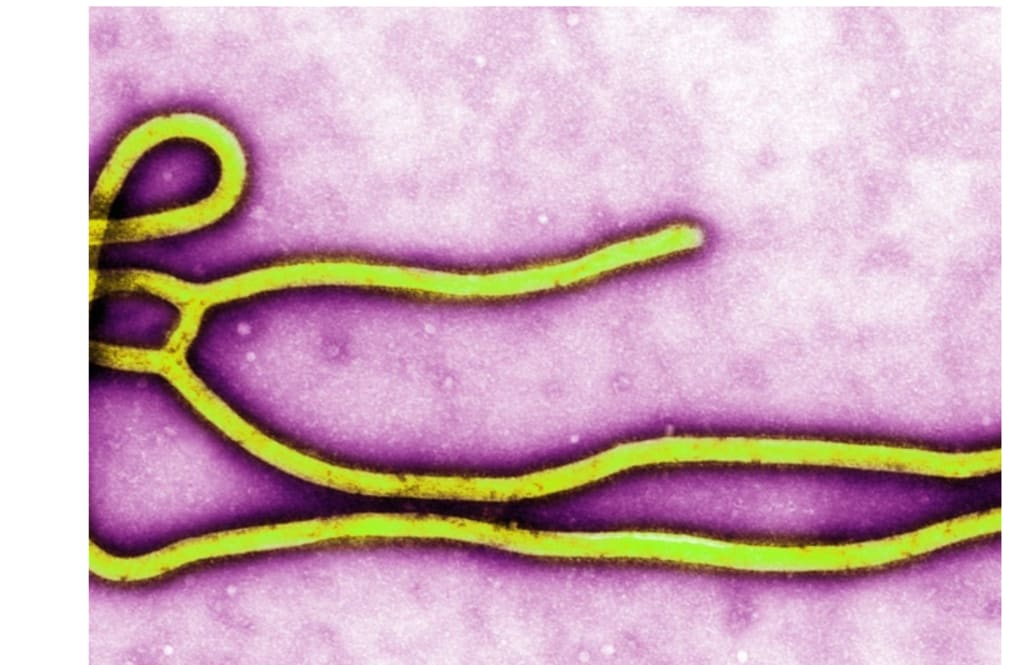
Even after being proclaimed cured, an Ebola survivor may still have the active virus in their brain for months. It can flare up again at times. It can occasionally rekindle an outbreak that everyone believed had ended.
Some survivors experience inflammation months after recovery, which can cause brain swelling or endanger their vision. Years after an outbreak ceased, it has resumed due to a single concealed virus. Until a researcher generated living human brain tissue in a dish, it was nearly impossible to see where the virus hides.
A bowl of brains
It's challenging to study Ebola inside the brain. The organ is protected by strong barriers that the immune system hardly ever checks, allowing invaders to proliferate. A living brain cannot be sampled in order to observe a virus in action.
At the Bernhard Nocht Institute for Tropical Medicine in Hamburg, Germany (BNITM), Lina Widerspick, Ph.D., worked on the issue. She managed to get around it with colleagues at the Icahn School of Medicine at Mount Sinai (ISMMS).
Cerebral organoids, which are pea-sized balls of living brain tissue, were created by the team using human stem cells. Surprisingly, these spheres resemble brains. They develop signal-carrying neurones, microglia, the immune cells that live in the brain, and astrocytes that nourish them.
Instead of determining the virus's path from a mouse, the scientists could observe how the virus would act in a human, not an animal, because the cells were human.
Ebola would not go.
After dripping Ebola onto the organoids, the crew bided their time. Over 90% of infections were successful. For the duration of the trial, which was 120 days, the virus then settled into a continuous, low simmer.
It wasn't dormant. The tissue continued to release new, contagious particles that may initiate a new infection during those months. This was not leftover detritus, but a live virus.
Another virus called Lassa responded completely differently, disappearing in a matter of weeks as though the tissue had. The close relatives of Ebola, such as the Marburg and Reston viruses, which do not even cause illness, dug in just as obstinately.
The brain's Ebola virus
Where it settled was the true surprise. Scientists debated whether Ebola could infect neurones at all for years. It is able to. Just six days after exposure, the virus's telltale signals appeared inside neurones, and they persisted for 120 days.
The virus infected the immune cells in the brain, which gathered around the diseased areas as if pulled in. Ebola has been discovered to persist in many of the same cells in a previous investigation conducted on monkeys.
The virus also infected other support cells. Since neurones were among the first cells damaged, The team believes that rather than being spectators caught up later, they might be an early footing.
An immunological standoff
The invader was not disregarded by the organoids. In an attempt to mobilise a defence, infected tissue released inflammatory alarm signals. However, it was insufficient.
As the months went by, the inflammation simply increased as the virus continued to multiply despite the reaction. That gradual burn is similar to what some survivors experience.
Some people experience eye inflammation that is severe enough to endanger their vision months after they recover, and one report discovered the live virus inside an inflamed eye.
Sometimes long after the initial disease, some people experience hazardous enlargement of the brain and its lining. The organoids demonstrated how that inflammation may be sustained by a covert infection.
Adapting to survive
After months of residing in the tissue, the virus evolved. Mutations in its genetic coding led to the emergence of new variations. Some copies were damaged and lacked components that they had to distribute independently.
Numerous modifications appeared to slow the virus rather than accelerate development. Ebola may be able to conceal itself by becoming slower and quieter, attracting less attention and using fewer resources.
A number of these mutations had never been found in surviving humans. The next question is whether they just happened to appear or if they aid in the virus's persistence.
What it reveals
No one has been able to observe Ebola surviving in living brain tissue until today. Once inside neurones and other brain cells, the virus produces infectious copies that fuel inflammation.
A senior author of the paper is Gustavo Palacios, Ph.D., a microbiologist at Mount Sinai. The organoids, in his opinion, are a beginning rather than an end. He stated, "More research is now necessary to examine the long-term interactions between virus and host."
Without relying so much on lab animals like mice and monkeys, the model gives researchers a human stand-in they have never had, allowing them to test antiviral medications and investigate how the virus hides.
The larger stakes are located outside the laboratory. According to one study, an outbreak was rekindled years after it ended due to a persistent infection in one survivor. Scientists may be able to prevent such from happening in the future with the use of tools that ultimately allow them to monitor the virus's hiding places.
About the Creator
Enjoyed the story? Support the Creator.
Subscribe for free to receive all their stories in your feed.
Comments
There are no comments for this story
Be the first to respond and start the conversation.