
His Brain Was 80. He Was 49. The Pills on His Nightstand Told the Rest.
A Shanghai man’s stroke led to a vascular dementia diagnosis. His MRI showed white matter damage. His blood pressure pills were still in the blister pack.
The Scan Did Not Match the Birthday
A 49-year-old man in Shanghai had a stroke. His MRI looked like an 80-year-old’s. The pills on his nightstand told the rest.
The chopsticks fell first. Then the right hand.
He was at dinner in Shanghai, 49 years old. He had high blood pressure. He stayed awake until two in the morning. He reached down with his right hand. The chopsticks slipped. He picked them up with his left hand. They fell again. His wife called 120.
At the hospital, the stroke team moved fast. A clot had blocked blood flow in his brain. He received thrombolysis. His hand came back. He could lift it. He could squeeze. The emergency was over.
Then a nurse said, “Close the window.”
He looked at the window. There was one window in the room. It was two feet away. He asked, “Which window?”
The scan
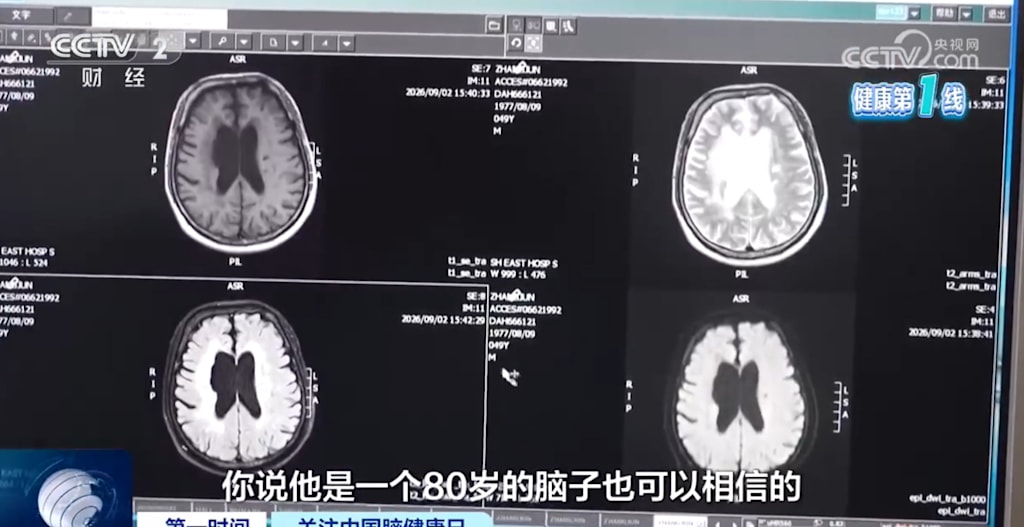
The MRI was done on a T2-FLAIR sequence. On that sequence, damaged white matter shows up bright. The report described extensive white matter hyperintensities around the ventricles. The radiologist pointed at the screen. “These areas should be dark,” he said.
They were not dark. They were white. Patchy at first, then confluent. The white matter tracts, the cables that connect one part of the brain to another, were injured. The brain itself was smaller than it should have been. At 49, his brain looked like the brain of an 80-year-old.
The diagnosis was vascular dementia. The cause was written in the small vessels.
What white matter hyperintensities mean
White matter hyperintensities are a footprint. They show that small arteries, arterioles, and capillaries in the brain have been damaged. The most common cause is high blood pressure.
When blood pressure stays high for years, the walls of these tiny vessels change. The endothelium, the inner lining, is injured. The blood-brain barrier becomes leaky. Proteins from the blood enter the brain tissue. Immune cells follow. Inflammation starts. The vessels that should deliver blood to the deep white matter cannot do their job. The tissue becomes chronically ischemic.
Oligodendrocytes, the cells that make myelin, are vulnerable to ischemia. Myelin wraps around axons like insulation around a wire. When myelin is lost, signals travel more slowly. When axons are damaged, some signals do not arrive at all.
This is why the man could not answer a simple instruction. The window was there. The word “window” was there. The connection between them was not.
Vascular dementia has a pattern. Executive function goes first. Processing speed drops. Attention wanders. Memory may be less affected than in Alzheimer’s disease. The man’s question, “Which window?”, was a speed problem. The brain was still receiving the instruction. It was just taking too long to route it.
Sleep was the multiplier
The news reports focused on his late nights. He had been sleeping from two in the morning until seven, maybe six hours, for years. That mattered. It did not matter alone.
A 2024 study in the Journal of the American Heart Association looked at 682 people from the Framingham Heart Study. It measured sleep duration and white matter hyperintensities. The finding was specific: short sleep was associated with higher white matter hyperintensity burden and worse executive function in people with hypertension. In people with normal blood pressure, the association did not hold.
The numbers were not huge, but they were consistent. In the hypertensive group, shorter sleep was linked to more white matter damage (β = −0.115, P = 0.042) and worse executive function (self-reported sleep: β = 0.041, P = 0.005; polysomnography: β = 0.045, P = 0.038). In the normotensive group, those links disappeared.
Hypertension had already weakened the pipes. Sleep loss turned up the pressure.
Sleep deprivation does several things at once. It raises sympathetic nervous system activity. Blood pressure does not dip at night the way it should. Morning blood pressure surges. Inflammation rises. TNF-α and other cytokines circulate. The blood-brain barrier, already damaged by hypertension, becomes more permeable.
Sleep loss also hits myelin directly. A 2026 study in PNAS found that sleep deprivation disrupts cholesterol metabolism in oligodendrocytes. Cholesterol is needed to build and maintain myelin. When that process fails, myelin function declines. Nerve signal conduction slows. Communication between brain regions becomes less synchronized. The paper linked this to deficits in cognition and movement.
There is also the glymphatic system. During sleep, the brain clears metabolic waste. Sleep loss impairs that clearance. Toxic proteins accumulate. Inflammation worsens. The white matter takes the damage.
The evidence was already there
The link between short sleep and dementia has been tracked for decades. A Nature Communications study followed nearly 8,000 people for about 25 years. At age 50 and 60, people who slept six hours or less had a higher risk of dementia than those who slept around seven hours. Those who slept short at 50, 60, and 70 had the highest risk. The association was not explained by other lifestyle factors.
A UK Biobank study using brain MRI found something similar. Compared with people who slept a medium amount, short sleepers had higher white matter hyperintensity loads, smaller cortical volumes, and worse white matter microstructure. The differences were visible on scans. They were not subjective complaints.
The man in Shanghai had both risk factors. He had untreated hypertension. He had chronic short sleep. The two combined.
Hypertension damages the small vessels. Sleep loss increases inflammation, disrupts blood pressure rhythms, and impairs myelin repair. The white matter is the meeting point. It is the tissue that gets hit from both sides.
The chain
His hypertension probably started years earlier. Maybe in his thirties. Maybe in his forties. He did not take medication. The pills sat on the nightstand in a blister pack. The foil was intact.
His sleep loss probably started around the same time. Work, screens, habit, whatever kept him awake until two. The body adapted. The blood pressure did not.
Over time, the small vessels narrowed and leaked. The white matter thinned. The connections slowed. At some point, a clot formed and blocked a vessel. The right hand stopped working. The thrombolysis cleared the clot. The hand moved again.
But the white matter was still damaged. The brain was still small. The question, “Which window?”, was still there.
The nightstand
After the stroke, he was moved to a regular ward. A nurse asked him to close the window. He looked at it. He did not ask which one this time. He stood up, walked over, and closed it. Then he sat back down.
On the nightstand beside the bed, the blood pressure pills were still in their blister pack. The foil had not been pushed through. The window was closed. The room was quiet.
The scan had shown the damage. The pills showed the years before it.
About the Creator
Jin
Writer of reamstories
https://reamstories.com/jin
Enjoyed the story? Support the Creator.
Subscribe for free to receive all their stories in your feed. You could also become a paid subscriber, letting them know you appreciate their work.
Comments
There are no comments for this story
Be the first to respond and start the conversation.