
When Development Ends, Does the Window Close With It?
A new study in adult mice suggests some consequences of altered brain development may remain biologically modifiable long after their origins are established.

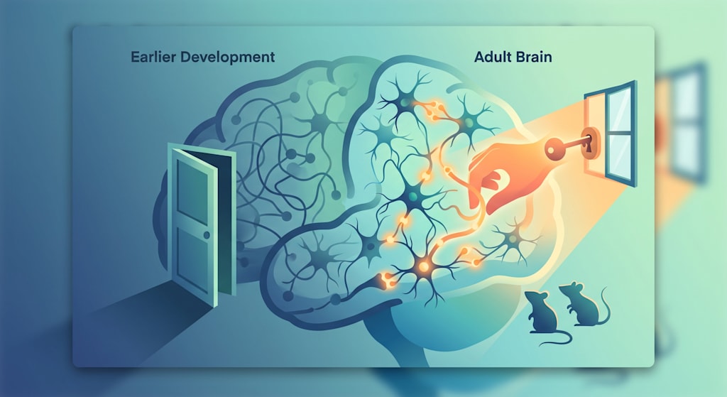
There is a quiet assumption buried inside how most of us think about the brain: that whatever happens during development becomes the brain's permanent architecture. A childhood of altered wiring hardens into an adult structure that can be managed or medicated around, but not fundamentally revisited. The early years are when the building gets built; afterward, you live in the house.
This assumption is not unreasonable. It draws on real biology. Critical periods exist; sensory systems, language acquisition, and aspects of social cognition all show heightened sensitivity to experience early in life, followed by a narrowing of that sensitivity as circuits mature. For disorders examined through a neurodevelopmental lens — including autism spectrum disorder, epilepsy, schizophrenia, and bipolar disorder — this question carries particular weight. If a developmental disruption set a trajectory in motion, and the adult brain has largely lost its capacity to be reshaped by experience, then whatever consequences persist into adulthood look less like an open problem than a settled one.
A new study published in Nature, led by researchers at Massachusetts General Hospital and Harvard Medical School, complicates this picture — not by claiming the brain can simply rewire itself, and not by proposing a treatment for autism, but by asking a narrower question. What if some of the biological mechanisms that would ordinarily let a circuit adjust to new experience are still present in the adult brain, just no longer working? And could restoring the mechanism itself — rather than undoing whatever went wrong in development — change what the circuit is capable of, even long after development has finished?
Inhibition Is Not an Off Switch
To understand why this question matters, it helps to reconsider what inhibitory neurons actually do. A common shorthand treats inhibition as a kind of brake — the neural equivalent of turning something off. That picture is misleading. A brain that only excited itself, without inhibition sculpting and timing that excitation, would not be a more active brain. It would be a brain incapable of representing anything specific, because every signal would blur into every other signal.
Inhibition is what gives neural activity its shape. In the hippocampus, a region central to memory and to spatial and social cognition, a class of fast-firing inhibitory cells called parvalbumin-expressing interneurons — PV neurons, for short — perform this shaping role with remarkable precision. Positioned within the circuitry connecting the dentate gyrus to the CA3 and CA2 subregions, PV neurons receive input from excitatory principal cells and feed inhibition back onto those same principal cells within milliseconds. This loop, known as feedforward inhibition, does not just dampen activity — it determines which populations of principal cells fire together as a coherent group, or "ensemble," encoding a specific experience, and which do not. Feedforward inhibition also helps generate sharp-wave ripples, brief bursts of coordinated network activity during rest and sleep thought to support memory consolidation. Excitation proposes; inhibition, in a sense, decides what gets registered as a distinct memory and what gets folded into noise.
For this system to work well, excitation and inhibition cannot simply coexist — they have to be coordinated, moment to moment and experience to experience. And that coordination depends on inhibitory neurons themselves having the capacity to change.
The Unusual Plasticity of PV Neurons
It might seem intuitive that only excitatory neurons — the cells that form and strengthen synapses in response to learning — need to be plastic. But the Nature study builds on prior work showing that PV interneurons themselves are dynamically responsive to experience. When an animal learns something new, or has a novel social encounter, its hippocampal PV neurons do not simply relay a fixed amount of inhibition. They adjust their own intrinsic excitability, restructure their synaptic connections to principal cells, and modify the strength and plasticity of those synapses in response to what the animal has just experienced — a process the researchers call experience-dependent PV interneuron plasticity, and treat as a distinct biological process worth investigating in its own right.
Prior work from this group had already shown that increased input from dentate gyrus mossy fibers — the axons of granule cells — onto PV neurons in CA3/CA2 triggers this kind of plasticity, strengthening feedforward inhibition and supporting spatial and social memory. What remained unknown was which genes, inside adult PV neurons, actually carry out this experience-triggered remodeling.
A Screen Built Around Real Experience
To find those genes, the researchers designed an input-specific translatome screen — a method for isolating and sequencing the messenger RNA being actively translated inside PV neurons, comparing a naive resting state to a state triggered by increased mossy fiber input. Rather than artificially forcing activity onto these neurons with electrical or chemical stimulation, the approach was built to mimic a physiological trigger, the kind of input change that ordinarily accompanies a real experience.
The results reached beyond the original question. A substantial number of the genes that turned on when PV neurons entered this experience-responsive state overlapped with genes already known, from human genetics, to be high-confidence risk factors for neurodevelopmental disorders — dozens of category 1 and S1 autism risk genes cataloged in the SFARI database, several genes implicated in schizophrenia through large-scale exome sequencing, and a set of bipolar disorder candidate genes. Roughly half of the autism-linked genes in this group were also independently associated with epilepsy, a pattern that tracks with how often seizures and cognitive impairment co-occur clinically.
The researchers are careful about what this overlap does and does not imply. It does not mean autism, schizophrenia, bipolar disorder, and epilepsy are the same condition, or share a common cause — these remain, individually, highly heterogeneous conditions with distinct genetic architectures and clinical presentations, and no mouse can model the lived complexity of any of them. What the overlap suggests instead is more specific: that genetically distinct developmental risk factors may converge, at the level of hippocampal circuitry, on the same underlying vulnerability — impaired capacity of PV neurons to change in response to experience. If that convergence holds up, it opens a strategic possibility: rather than needing a separate intervention for every risk gene, it might eventually be possible to target the shared downstream process those genes feed into.
Why Meis2
Among the candidate genes, the researchers focused on Meis2, a homeobox transcription factor with a well-established developmental role — it helps specify the identity of certain inhibitory projection neurons during embryonic life. In the adult hippocampus, Meis2 is normally expressed only at very low levels in PV neurons, and its function there had not been characterized. When the researchers artificially increased Meis2 expression in PV neurons of otherwise healthy adult mice, using enhancer-driven viral vectors targeting PV cells with high specificity, it increased the density of PV neuron synapses onto principal cells in CA3/CA2. Other candidate genes from the screen — Tbr1, Bcl11a, and Herc1 — produced similar increases, suggesting several converge on synapse formation, but Meis2 was chosen for deeper investigation as a proof-of-concept case.
The choice of animal model mattered. The researchers used Cntnap2 knockout mice, a well-established genetic model in which mice lack a gene strongly linked to autism spectrum disorder in humans, and which develop neuronal migration abnormalities, hyperexcitability, loss of PV interneurons, cognitive impairments, and, by around six months of age, spontaneous seizures. The authors state directly that their goal was not to model autism specifically, but to use a well-characterized neurodevelopmental risk model — one whose deficits are established early and persist — as a testbed for a broader question about circuit plasticity.
What Happened When Meis2 Was Restored, in Adulthood
The Cntnap2-deficient mice used in this study were adults, roughly two months old, by the time any intervention occurred — well past the developmental window in which their circuit deficits originated. At baseline, these mice had fewer PV neuron synapses onto CA3/CA2 principal cells than wild-type littermates, and, notably, they had lost something wild-type mice retained: the ability of a novel social encounter to trigger new PV synapse formation and rising Meis2 expression in the first place. The deficit was not just structural. It was also a lost capacity to respond to experience at all.
Restoring Meis2 expression specifically in CA3/CA2 PV neurons of these adult knockout mice — verified using two independent genetic strategies — reversed a striking range of downstream measures. Perisomatic PV synapses onto principal cells were restored. The abnormal excitation-inhibition balance onto CA2 principal cells, tilted toward excess excitation in untreated knockout mice, was corrected. The intrinsic excitability of the PV neurons themselves, which had been blunted, was restored. A specific form of inhibitory synaptic plasticity — normally triggered by patterned mossy fiber stimulation, and essentially absent in untreated knockout mice — was reinstated.
These circuit-level changes were accompanied by improvements in cognition. In a task assessing memory for object locations, and in a task assessing social recognition, knockout mice with restored Meis2 performed comparably to wild-type mice, while untreated knockout mice did not. An activity-tagging technique showed that restoring Meis2 increased the specificity with which the same neuronal ensemble was reactivated upon a later, related social experience — a plausible mechanistic link between improved circuit function and improved memory. Recordings taken during rest additionally showed that sharp-wave ripple activity, diminished in untreated knockout mice, was restored toward wild-type levels.
Perhaps the most clinically resonant finding involved seizures. Months later, when mice were old enough for spontaneous seizures to typically emerge in this model, seizures were detected in 10 of 14 untreated knockout mice, compared with 3 of 12 mice that had received the Meis2 intervention during two weeks of continuous recording; seizure frequency was also markedly reduced among the treated animals. Wild-type mice given the same intervention showed no comparable increase in seizures — a relevant safety signal within this single preclinical study, though far short of anything resembling a human safety profile.
The Adulthood Question
It would be easy to read all of this and conclude that a form of developmental damage was undone. That is not quite what the data show, and the distinction is not pedantic. The researchers did not intervene during development, and they did not alter the animals' genotype or reverse the original abnormality that produced the knockout phenotype. Cntnap2 remained absent throughout the animals' lives. What changed was a single downstream mechanism, in a specific cell type, in a specific hippocampal circuit, well after that circuit had already matured under abnormal conditions.
This is the conceptual core of the study. There is a difference between correcting a developmental process while it is unfolding and restoring, later, the mature circuit's capacity to still respond to experience the way a healthy circuit would. The first strategy intervenes upstream, before deficits are locked in. The second accepts that a downstream consequence already exists and asks whether the circuit retains some latent machinery for adjusting itself regardless. That the second strategy worked here suggests at least some of what looks like a fixed consequence of early development may be a mechanism sitting dormant rather than a structure permanently lost. Restoring the mechanism, rather than rewinding the history that damaged it, was sufficient to change several downstream outcomes.
This is a genuinely different claim from saying the brain "rewired itself." Nothing here suggests global structural transformation or a general loosening of adult plasticity. A single gene, in a defined population of inhibitory neurons, in one hippocampal circuit, was manipulated, and specific, measurable properties of that circuit changed as a result. The findings are precise rather than sweeping, and their significance lies in that precision.
What This Study Does Not Show
Given how easily findings like these get inflated in translation, it is worth stating plainly what this research does not establish.
It was conducted entirely in mice, using genetically engineered viral tools delivered directly into the brain — a method with no current equivalent as a safe, approved human intervention. It does not demonstrate a treatment for autism, epilepsy, schizophrenia, bipolar disorder, or any other neurodevelopmental condition in people. Increasing Meis2 expression in a defined population of hippocampal neurons in a mouse is not equivalent to having identified a therapy; the delivery method used here would require extensive further work on targeting precision, dosing, long-term safety, and durability before human translation could reasonably be considered, and most candidates that look promising in a single mouse model do not survive that process. Mouse behavioral assays, however carefully designed, capture narrow facets of cognition and social behavior; they cannot capture the breadth of what autism, or any of these other conditions, means as a human experience across a lifetime. Restoring select circuit-level properties in one hippocampal pathway is not equivalent to reversing an entire neurodevelopmental condition, which involves many brain regions, developmental stages, and individual differences this study did not address. And a single mouse model, built around a single gene, cannot establish that the same mechanism operates the same way across the genetically and biologically diverse conditions loosely gathered under the neurodevelopmental disorder umbrella — even where the genetic overlap identified in the screen is suggestive.
None of this diminishes what the study accomplished. It simply keeps the finding in its proper register: a carefully controlled proof of concept, in one circuit, in one model, that raises a testable scientific possibility rather than settling a clinical question.
Rethinking the Therapeutic Window
What this study changes is not what can currently be offered to any patient, but how researchers might reasonably think about where the boundaries of biological possibility sit. The conventional framing of a therapeutic window assumes that once a critical developmental period closes, opportunities for meaningful circuit-level intervention close with it, leaving only management of a fixed condition. This study suggests that assumption may be too coarse. Some downstream consequences of an early developmental disruption may persist not because the original disruption is irreversible, but because a maintenance mechanism — the capacity of a specific cell type to keep responding to experience — has itself become impaired and is, in principle, still modifiable in adulthood.
That is a scientific hypothesis suggested by this work, not a general law it has proven. It will need testing across other models, other circuits, and eventually, cautiously, other species before it means anything for how neurodevelopmental disorders are approached clinically. But it offers a genuinely different way of asking the question. Rather than asking only what went wrong during development, it becomes possible to ask what the adult circuit, as it currently stands, still retains the capacity to do — not because development can be undone, but because permanence and origin are not necessarily the same thing. A disruption can begin early in life while the mechanisms sustaining its consequences remain, in some circuits, open later than assumed.
References / Further Reading
Shih, Y.-T., Alipio, J. B., Klaft, Z.-J., Green, N., Mohapatra, A. N., Goode, T. D., Panchanatham, M., Pathak, D., Wong, L. P., Sadreyev, R., Hyun, J. H., Ahmed, O., Dulla, C. & Sahay, A. "Procognitive restoration of PV neuron plasticity in neurodevelopmental disorders." Nature (2026). DOI: 10.1038/s41586-026-10907-8
Peñagarikano, O., Abrahams, B. S., Herman, E. I., Winden, K. D., Gdalyahu, A., Dong, H., Sonnenblick, L. I., Gruver, R., Almajano, J., Bragin, A., Golshani, P., Trachtenberg, J. T., Peles, E. & Geschwind, D. H. "Absence of CNTNAP2 leads to epilepsy, neuronal migration abnormalities, and core autism-related deficits." Cell 147, 235–246 (2011). DOI: 10.1016/j.cell.2011.08.040
Su, Z., et al. "Dlx1/2-dependent expression of Meis2 promotes neuronal fate determination in the mammalian striatum." Development 149, dev200035 (2022). DOI: 10.1242/dev.200035
Shih, Y.-T., Alipio, J. B. & Sahay, A. "An inhibitory circuit-based enhancer of DYRK1A function reverses Dyrk1a-associated impairment in social recognition." Neuron 111, 3084–3101 (2023). DOI: 10.1016/j.neuron.2023.09.009
Contractor, A., Ethell, I. M. & Portera-Cailliau, C. "Cortical interneurons in autism." Nature Neuroscience 24, 1648–1659 (2021). DOI: 10.1038/s41593-021-00967-6
About the Creator
Khali Sollis
Khali Sollis is a writer and independent researcher exploring the science of the human mind and behavior. Her work examines questions at the intersection of neuroscience, psychology, cognition, mental health, and everyday human experience.
Enjoyed the story? Support the Creator.
Subscribe for free to receive all their stories in your feed.
Comments
There are no comments for this story
Be the first to respond and start the conversation.