
A Calorie Is a Measurement. A Molecule Is a Message.
Two sugars with identical caloric value speak to the brain through different nerves entirely — and a 2026 mouse study on hunger neurons suggests that "energy" and "information" are not the same thing to a nervous system.

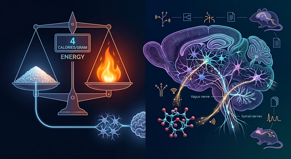
A calorie is a unit of energy. Nutrition labels don't arrive at that number by incinerating food in a lab; modern food energy values are typically calculated using metabolizable-energy systems, such as Atwater factors, that estimate how much of a nutrient's raw energy content the body can actually extract and use. Either way, the number is a quantitative measure of available fuel. Glucose and fructose are even the same chemical formula, C₆H₁₂O₆, just arranged differently, and both supply roughly the same number of calories per gram. If you replaced one with the other in a recipe, the calorie count on the label wouldn't change by a single digit.
But a body doesn't process food only by asking how much energy it contains. The body has its own instruments for finding out what has arrived in the gut: transporters, hormones, sensory nerves, and neurons that specialize in detecting exactly this kind of thing. Those instruments do not necessarily read glucose and fructose the same way, even when an energy-accounting method would call them equivalent. A study published in June 2026 gives an unusually precise look at just how differently two calorically matched sugars can be handled once they leave the plate and enter a body that is trying to figure out what it just ate.
A Theory About Hunger Neurons, Tested
Deep in the hypothalamus sits a small population of neurons that neuroscientists have spent the last two decades mapping in detail: agouti-related protein neurons, or AgRP neurons. When these neurons are activated, they strongly promote feeding behavior; their activity is also modulated by nutrients reaching the gut, dropping within minutes of ingestion in a way that precedes full digestion [1]. Their activity is not driven by post-ingestive signals alone — sensory cues associated with food, such as the sight or smell of it, can shift AgRP activity even before anything is swallowed — but the post-ingestive, gut-driven component of that signal is the one this study examines. Because that gut-driven drop-off happens so quickly after eating, researchers have suspected that AgRP neurons function, at least in part, as a rapid-response sensor wired directly into gut signals rather than one that simply waits for nutrients to show up in the bloodstream.
One open question was whether the size of this gut-driven AgRP response tracks calories as an undifferentiated quantity — whether a calorie from any source turns the signal down by roughly the same amount. A team led by Amber Alhadeff at the Monell Chemical Senses Center and the University of Pennsylvania tested that question directly, by comparing how AgRP neurons in mice responded to equal caloric doses of glucose versus fructose [1]. This is worth stating plainly and keeping in view throughout: the experiment was conducted in mice, using intragastric or intestinal infusions and fiber-photometry recordings of neural activity, not in humans, and its findings describe mouse neurobiology first and foremost.
The result cut against the simple "calorie is a calorie" model of AgRP signaling. Glucose suppressed AgRP neuron activity substantially. Fructose, delivered in an equicaloric amount, suppressed it far less [1]. High-fructose corn syrup — the common food additive that blends the two sugars — fell in between, suppressing AgRP activity more than fructose alone but less than pure glucose, roughly in proportion to its glucose content [1]. Total short-term food intake in the mice didn't differ much between the two sugars; the animals ate similar amounts regardless of which sugar they'd been given. But when the mice were offered a choice between two flavored liquids, one sweetened with glucose (or the glucose-fructose blend) and one with fructose alone, they consistently favored the glucose-containing option [1]. The researchers interpret this as evidence that the size of the AgRP response is not just a passive readout of calories consumed; it appears to help construct preference itself, independent of how much was eaten in that particular sitting.
Two Different Nerves for Two Different Sugars
The more striking part of the paper is mechanistic. The Alhadeff lab's earlier work had already shown that different macronutrients don't share a single delivery route to AgRP neurons: fat signals through the vagus nerve, while glucose signals primarily through spinal, or splanchnic, sensory afferents that run from the gut and liver up through the spinal cord rather than through the vagus [3]. The new study asked where fructose fits into this map, and the answer was that it doesn't fit into either category the way expected. Fructose triggered a distinctly different pattern of vagal nerve cell activity than glucose did, and when the researchers surgically interrupted vagal signaling between the gut and brain, glucose was still able to suppress AgRP activity through its spinal route — but fructose's already-modest effect was lost [1]. Mechanistically, the team traced this to the gut hormone peptide YY (PYY): fructose consumption caused a larger and more sustained rise in circulating PYY than glucose did, and this PYY appears to act on Y2-receptor-expressing vagal neurons to deliver fructose's signal to AgRP neurons [1]. That elevated PYY response to fructose relative to glucose has also been observed directly in humans: in a 2015 study using paired, equicaloric glucose and fructose drinks in 24 healthy volunteers, plasma PYY specifically at the 60-minute mark was higher after fructose than after glucose, even though overall PYY exposure across the session — measured as area under the curve — did not differ significantly between the two drinks [2]. That narrower, timepoint-specific human finding is one of the few points in this line of research where mouse and human data actually converge.
So the two sugars aren't just producing different-sized effects on the same wire. They appear to be using two different wires: glucose primarily via the spinal pathway established in the lab's earlier work, fructose via a vagal route gated by a gut hormone. That a single downstream neuron population can be reached by two structurally distinct nerve pathways, carrying different chemical envoys, for two sugars with identical energy content, is the detail that gives this study its conceptual weight.
Energy Value and Biological Information Are Not the Same Property
It is tempting to describe this finding as proof that "fructose fools the brain" or that "not all calories are equal," and both phrases are worth resisting, because they compress a subtler point into a slogan. By standard energy-accounting measures, the calories are equal: a gram of fructose and a gram of glucose supply the same available energy. What this study shows is that equal caloric value does not imply identical neural signaling. Fructose and glucose are absorbed by different intestinal transporters and processed by different enzymes once inside cells — fructose is preferentially phosphorylated by an enzyme called ketohexokinase, entering a metabolic pathway distinct from the one glucose enters [6] — and it is plausible that these upstream differences in transport and processing are part of what allows the two sugars to provoke different hormonal and neural signals downstream, as this study found. The point this article rests on is narrower than a claim about overall metabolic equivalence: it is that the information a nutrient generates in transit — which transporters absorb it, which hormones its presence provokes, which nerve fibers are built to detect those hormones — is a separate property from how much energy it yields, and the two properties don't have to move together.
This distinction — between a substance's energy content and the biological signal it generates on the way to being used — has a longer history than this one paper. Work using mice that lack functional sweet-taste receptors showed, back in 2008, that animals could still develop a preference for calorie-containing sugar solutions over non-caloric ones, purely from post-ingestive effects that had nothing to do with taste [4]. That is, the reward value of a nutrient can be constructed after it is swallowed, using information gathered from inside the gut rather than from the tongue. And in the years since, researchers have found that specialized gut cells, sometimes called neuropod cells, form direct synaptic connections with vagal nerve fibers, allowing luminal contents to be reported to the brain with a speed and precision that a leisurely, diffuse hormone release could not achieve on its own [5]. Together, these lines of research support a picture of the digestive tract as something closer to a sensory organ than a purely mechanical processing tube — one equipped with receptors, transporters, and nerve endings whose job is to identify, not just break down, what has been eaten.
None of this requires abandoning the calorie as a useful unit. It is still the right tool for asking how much energy a food will contribute to the body's overall balance sheet. It is simply not the right tool for asking how the nervous system will represent that food, because "how much energy" and "what kind of molecule, detected by which pathway" turn out to be separable questions, at least for the neurons this study examined.
What This Study Does Not Show
It is worth being explicit about the boundaries of this evidence, because the temptation to over-extend a finding like this one is real. The study does not show that fructose causes overeating, weight gain, or addiction; short-term food intake in the mice was similar regardless of which sugar they consumed, and the paper doesn't address long-term metabolic outcomes at all [1]. It does not establish that AgRP neurons behave this way in humans, because there is currently no way to record from individual hypothalamic neurons in a living human being; the parallel human evidence here is limited to hormone measurements, specifically the elevated PYY response to fructose that has independently been observed in people, not to neural activity itself [2]. It also says nothing about whole fruit, where fructose arrives bound up with fiber, water, and a very different absorption profile than a purified fructose solution or a sugar-sweetened beverage — this study used isolated sugars, not whole foods, and the two should not be treated as biologically interchangeable. What the study does establish, with reasonable confidence for the species and conditions tested, is that a specific population of hunger-regulating neurons in mice is more strongly inhibited by glucose than by an equal number of calories from fructose, that this difference tracks with beverage preference rather than short-term intake, and that the two sugars reach those neurons via distinguishable neural routes.
Reading Food as a Body Reads Food
There is a reason it feels strange, on first encounter, to hear that two calorically identical sugars might not mean the same thing to the brain. We tend to think of eating as a transaction in energy: food goes in, energy comes out, and the conversion rate is fixed by chemistry. That framework isn't wrong, exactly. It's incomplete, because it describes food only from the outside, the way an energy-accounting formula does, and says nothing about what happens once a body starts asking its own questions about the substance passing through it — which transporter grabs it, which hormone it provokes, which nerve carries the news upstairs.
The calorie, though, is a fairly recent invention — a bookkeeping unit that nutrition science built in order to compare foods on a single scale. AgRP neurons, spinal afferents, vagal nerve fibers, PYY, ketohexokinase: none of these were designed with that scale in mind. They evolved over a much longer and more indifferent timescale, shaped by a very different problem — not totaling up energy, but telling one molecule apart from another quickly enough to matter for survival. The nervous system was never trying to answer the question a nutrition label answers. It was answering a different question entirely: not simply how much came in, but what came in, and by which route it announced itself. A calorie count and a hunger neuron are both, in their own way, forms of measurement — they simply weren't built to measure the same thing.
References
[1] McKnight, A.D., de Araujo, A., Hsu, F.Y., Vargas-Elvira, A.G., Acosta, A.A., Smith, M.M., Iwueze, W., de Lartigue, G., & Alhadeff, A.L. (2026). Attenuated hypothalamic response to fructose via a dedicated gut-brain pathway. Neuron. Published online June 10, 2026. DOI: 10.1016/j.neuron.2026.05.013.
[2] Luo, S., Monterosso, J.R., Sarpelleh, K., & Page, K.A. (2015). Differential effects of fructose versus glucose on brain and appetitive responses to food cues and decisions for food rewards. Proceedings of the National Academy of Sciences, 112(20), 6509–6514. DOI: 10.1073/pnas.1503358112.
[3] Goldstein, N., McKnight, A.D., Carty, J.R.E., Arnold, M., Betley, J.N., & Alhadeff, A.L. (2021). Hypothalamic detection of macronutrients via multiple gut-brain pathways. Cell Metabolism, 33(3), 676–687.e5. DOI: 10.1016/j.cmet.2020.12.018.
[4] de Araujo, I.E., Oliveira-Maia, A.J., Sotnikova, T.D., Gainetdinov, R.R., Caron, M.G., Nicolelis, M.A.L., & Simon, S.A. (2008). Food reward in the absence of taste receptor signaling. Neuron, 57(6), 930–941. DOI: 10.1016/j.neuron.2008.01.032.
[5] Kaelberer, M.M., Buchanan, K.L., Klein, M.E., Barth, B.B., Montoya, M.M., Shen, X., & Bohórquez, D.V. (2018). A gut-brain neural circuit for nutrient sensory transduction. Science, 361(6408), eaat5236. DOI: 10.1126/science.aat5236.
[6] Hannou, S.A., Haslam, D.E., McKeown, N.M., & Herman, M.A. (2018). Fructose metabolism and metabolic disease. Journal of Clinical Investigation, 128(2), 545–555. DOI: 10.1172/JCI96702.
The original 2026 findings discussed here were first brought to public attention by the National Institutes of Health's NIH Research Matters, "Fructose and glucose trigger different brain responses" (July 14, 2026), which summarized the McKnight et al. Neuron paper; that NIH summary served as the prompt for this essay but is not cited as a source for scientific claims, which point to the primary literature above.
About the Creator
Khali Sollis
Khali Sollis is a writer and independent researcher exploring the science of the human mind and behavior. Her work examines questions at the intersection of neuroscience, psychology, cognition, mental health, and everyday human experience.
Enjoyed the story? Support the Creator.
Subscribe for free to receive all their stories in your feed.
Comments
There are no comments for this story
Be the first to respond and start the conversation.