
Exploring the Relationship Between Aphantasia and Dimethyltryptamine Levels in Cerebrospinal Fluid
Looking Inward

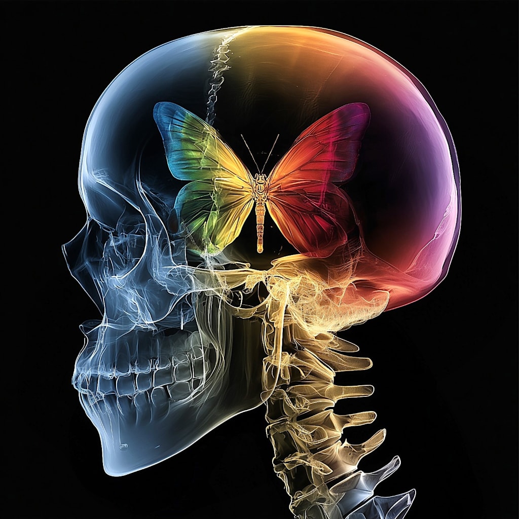
For the purposes of this exploration, let us define a hallucination as the experience of something that is only present in one’s mind. The hallucinogenic substance known as dimethyltryptamine, or DMT, enables the vivid experience of things that are only present in one’s mind. Our imagination also enables us to experience things that are only present in the mind. Studies report that low concentrations of endogenous DMT (that is, DMT which forms naturally inside one’s body) between 0.12 to 100 ng/ml are normally present in human cerebrospinal fluid [1]. Aphantasia is the inability to picture things in one’s “mind’s eye,” or imagination. There are two types of aphantasia: congenital aphantasia, which is present from birth, and acquired aphantasia, which develops through brain injury or illness. Studies show there are no significant structural differences between the brain of someone with congenital aphantasia and the brain of someone who can visualize things in their mind’s eye, so the primary contributor to congenital aphantasia must be a matter of functional divergence as opposed to structural abnormality. To be clear, there are some subtle structural differences in aphantasic brains, but not enough to explain the disruption of the visualization process [2]. This paper proposes that extremely low levels of endogenous DMT in human cerebrospinal fluid (near or below the lower end of the reported range, or ≲ 0.12 ng/ml) may contribute to the functional differences underlying congenital aphantasia.
The validity of this hypothesis can be easily determined through the extraction of cerebrospinal fluid from a subject with a vivid imagination (as determined by a high score on the Vividness of Visual Imagery Questionnaire, or VVIQ) and a subject with extreme aphantasia, i.e., the inability to visualize anything whatsoever in their mind’s eye (also determined by the Vividness of Visual Imagery Questionnaire). Extracting a sample of cerebrospinal fluid is commonly known as a spinal tap. It’s a standard medical procedure that takes about 30 minutes, with the actual collection of fluid only taking a few minutes. I think 25 subjects with vivid imaginations and 25 subjects with extreme aphantasia would initially be enough to indicate whether or not a relationship exists between endogenous DMT levels in cerebrospinal fluid and aphantasia. Cerebrospinal DMT levels can be measured using liquid chromatography-tandem mass spectrometry. I myself do not have the means to conduct such research, but I have made this information freely available through several public sources for anyone who might.
If an obvious difference in cerebrospinal DMT levels between the aphantasic group and the control group is found, this would justify a larger, more in-depth study. If no meaningful difference is found, this would suggest that the underpinnings of congenital aphantasia might be psychological rather than physical.
Approximately 2% of the population is aphantasic [3]. The term aphantasia was coined by Adam Zemam and his associates in 2015 [4]. Francis Galton, a 19th-century psychologist, devised the earliest method of measuring the vividness of imaginative visualizations. His “breakfast table questionnaire” asked participants to rate the “illumination, definition and coloring of your breakfast table as you sat down to it this morning.” The test revealed that some participants had no ability to visualize things in their mind’s eye [5].
The confirmation of a correlation between endogenous DMT and imaginative visualization which indicated that different levels of endogenous DMT in cerebrospinal fluid result in different degrees of coherency and vividness regarding imaginative visualization would furthermore explain why there are varying degrees of aphantasia [6].
Visualizing something in your mind’s eye involves a combination of frontoparietal regions and areas involved with memory, including the default mode network, along with the fusiform and primary visual cortices [7]. The process of mental visualization has been described as “vision in reverse.” When mentally visualizing something, the usual flow of information from the retina to multimodal cortices is reversed, utilizing extensive feedback pathways that play a less prominent role in vision [8]; however, the neural correlates for the vividness of mental imagery are controversial. As it stands, the vividness of mental imagery is associated with the degree of activation in higher-order visual areas and regions of the limbic system [9], but there is no current explanation for the degree of activation itself, unless my hypothesis is correct.
Multiple studies have shown that psychedelics increase neuroplasticity and change functional dynamics in the visual cortex [10]. DMT alters the visual system by increasing communication with associative networks, weakening regional boundary constraints, and amplifying feedback signals, making the visual cortex more sensitive to internally generated imagery. This implies that unusually low concentrations of cerebrospinal DMT could potentially result in inhibited interregional communication and duller feedback signals, making the visual cortex less responsive to internally generated imagery.
Lending support to my hypothesis, Dos Santos and colleagues describe a case of ayahuasca usage by a man with aphantasia, the first such report of someone with aphantasia using a hallucinogen (2018). The subject reported “an improvement in their visual imagery following one particular instance of ayahuasca use,” indicating a relationship between aphantasia and dimethyltryptamine [11].
References
1. Riba, Jordi, et al. “Metabolism and Disposition of N,N-Dimethyltryptamine and Harmala Alkaloids after Oral Administration of Ayahuasca.” Drug Testing and Analysis, vol. 4, 2012, p. 610. doi:10.1002/dta.1344.
2. Takamura, Yusaku, et al. “Fronto temporal Structural Alterations in Congenital Aphantasia.” bioRxiv, 2026, https://doi.org/10.1101/2026.03.22.713248.
3. https://pmc.ncbi.nlm.nih.gov/articles/PMC8186241/
4. Gallagher, James. “Aphantasia: A Life without Mental Images.” BBC News, 26 Aug. 2015. Archived 26 Aug. 2015. Accessed 26 Aug. 2015.
5. Galton F. 1880. Statistics of mental imagery. Mind. 5:301–318.
6. “Aphantasia Explained: What It Is, How the Mind Works Without Mental Images, and Why It’s Not a Disorder.” Ummeed Rehab, www.ummeedrehab.com/aphantasia-explained-mind-without-mental-images/ Accessed 27 Apr. 2026.
7. Winlove, C. I. P., et al. “The Neural Correlates of Visual Imagery: A Co ordinate Based Meta analysis.” Cortex, vol. 105, 2018, pp. 4–25.
8. Breedlove, J. L., et al. “Generative Feedback Explains Distinct Brain Activity Codes for Seen and Mental Images.” Current Biology, vol. 30, no. 12, 2020, pp. 2211–2224.e6. doi:10.1016/j.cub.2020.04.014.
9. Fulford, J., et al. “The Neural Correlates of Visual Imagery Vividness—An fMRI Study and Literature Review.” Cortex, vol. 105, 2018, pp. 26–40.
10. Lepow, Lauren, Hitoshi Morishita, and Rachel Yehuda. “Critical Period Plasticity as a Framework for Psychedelic Assisted Psychotherapy.” Frontiers in Neuroscience, vol. 15, 2021, article 710004.
11. Luke, David. “Reply to ‘Ayahuasca Turned on My Mind’s Eye’: A Case of Acquired versus Congenital Aphantasia, as Evidenced with DMT Use?” Journal of Psychedelic Studies, vol. 2, no. 2, 2018, pp. 97–98. doi:10.1556/2054.2018.014.
About the Creator
J. Martin Strangeweather
J. Martin Strangeweather is a poet, novelist, journalist, artist, teacher, philosopher, thanatologist, and psychopomp. He founded the Santa Ana Literary Association in 2020, and originated the eidetic afterlife hypothesis in 2024.
Enjoyed the story? Support the Creator.
Subscribe for free to receive all their stories in your feed.
Comments
There are no comments for this story
Be the first to respond and start the conversation.