
Electrocardiography (ECG/EKG) - basics
Electrocardiography (ECG/EKG) - basics

An electrocardiogram or ECG is a device that uses electricity to visualize the heart's electricity. By watching the waves of depolarization during each heartbeat, an ECG can show how the heart is working. The way it looks depends on the type of electrodes you're using, with lead II typically being used on one side of the body and lead III on the other. For this particular example, see this big positive deflection?
When studying the heart, it's important to be aware of the basic principles by starting with an example. With only one pair of electrodes, we can look at the heart in different ways. Remember that when cells are at rest, they are negatively charged relative to their surroundings and when they depolarize, they become positively charged.
Slightly negative charge in the external environment.
So let's say when this group of cells is at rest, they're red, and turn around
They appear green when depolarizing waves pass through them.
Now, if we freeze this "depolarization wave" as it travels through the cell, half
cells are positive or depolarized, half are negative or dormant, so there is
The difference in charge between the batteries in the set.
You can think of the charge difference as a dipole since there are two electric dipoles
pole, we can draw this dipole as an arrow or vector pointing to the positive
Charging, remember the electrodes detect the charge on the outside of the battery, so this
Point to where the positive charge is on the outside.
If the dipole vector is now pointing positively, the ECG
The records show it is positively deflected - the larger the dipole, the greater the deflection.
If we keep pausing, everything will depolarize, no difference
Responsible, now there are no dipoles and therefore no deflection.
Moments later, the repolarized wave passes through.
Pausing again midway, the vector dipole now moves in the opposite direction, and
Facing the negative electrode, which means there is a negative deflection
ECG recording.
Likewise, the larger the dipole, the larger the negative deflection.
While it would be nice if the depolarization waves were perfectly aligned with the electrodes,
Usually this is not the case, so let's look at the final vector components
parallel to the electrode.
For example, suppose depolarization happens like this - at an angle, then we would have
Split the vector into two parts.
One is parallel to the electrodes and one is perpendicular.
We're dealing with the one that goes to the positive pole.
causes a deflection, but since this arrow is shorter, it causes a slightly smaller deflection
Distraction than before, i.e. the degree of distraction in the ECG recording
always corresponds to the magnitude or magnitude of the dipole in the direction of the electrode.
The vertical components do not point towards the electrodes, so they do not cause their
diversion.
In fact, if there is a vertically upward depolarization wave
to the positive and negative electrodes, there is no deflection at all!
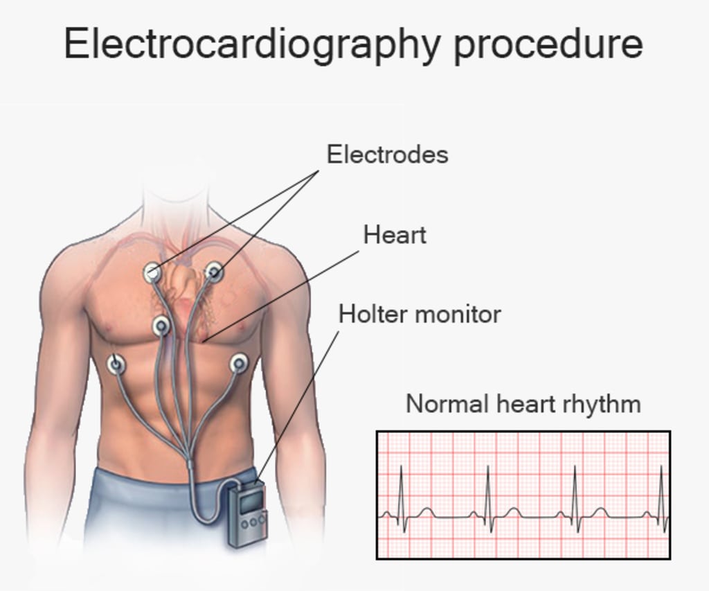
In a standard ECG there are 10 electrodes - four limb electrodes, one on the left arm,
Right arm, left and right leg and six precordial electrodes - V1 to V6
or the level of the heart, and each electrode is arranged to detect any wave
Positive charges are coming towards them, which, based on what we already know, means they are
positive.
These are collectively called the chest leads.
Meanwhile, on the coronal plane, non-neutral leads are represented as extended right vectors or
aVR on the right arm and Augmented Vector Left or aVL on the left arm - both
Expressed as a vector 30 degrees above the horizon.
Finally, the left foot has the Augmented Vector Foot or aVF, which is anatomically
Not straight down, but close enough to end up representing the vector direction
directly on the chart.
Like the precordial electrodes, aVR, aVL and aVF detect any positive deflection
coming towards them.
Now, in addition to these three limb leads, there are also bipolar limb leads called lead
1, 2, and 3, which are recorded using two electrodes instead of just one.
Lead 1 uses the Right Arm as the negative pole and Left Arm as the positive pole, forming
a vector that goes to the right.
Lead 2 uses the right arm as the negative pole and the left leg as the positive pole,
forming a vector that goes to the +60 degree mark.
And lead 3 uses the left arm as the negative pole and the left leg as the positive pole,
forming a vector that goes to the +120 degree mark.
So, in total you’ve got 6 leads from the limb leads, 6 from the chest leads, leading
to a grand total of 12, which gives you your 12-lead ecg.
Now the point of having all these leads is to get different views of the heart.
Making it easier to see exactly how the wave of depolarization moves through the heart.
As an example, consider how the 6 chest leads - V1 through V6 - register this depolarization
wave form called the QRS complex.
The exact same depolarization wave might appear mostly negative in V1 and V2, isoelectric
in V3, and mostly positive in V4, V5, and V6, all because of the exact direction and
magnitude of the vectors at different points in time.
Similarly, each limb lead provides a unique perspective of the depolarization wave. The limb leads and chest leads can be categorised based on the heart regions they are closest to, and issues in specific leads or groups of leads may indicate a particular area of the heart affected by disease. Leads II, III, and aVF are "inferior" leads as they are near the inferior heart wall that receives blood from the right coronary artery. Leads I and aVL, along with chest leads V5 and V6, are "lateral" leads and are near the lateral heart wall that receives blood from the left circumflex artery. V1 and V2 are "septal" leads as they are closest to the interventricular septum, while V3 and V4 are "anterior" leads as they are nearest to the anterior heart wall. Both the septal and anterior regions are served by the left anterior descending artery. To summarise, a standard ECG has ten electrodes - four limb electrodes and six precordial electrodes that wrap around the chest.
These specific electrodes are utilised to produce 12 leads, with each lead demonstrating the respective movement in question.
Heart cells have a positive charge on their outer surfaces.
The ECG reading displays a positive depolarization wave moving towards one of the electrodes.
A deflection can be described as a phenomenon in which one object moves towards another, and the other object moves away as a negative deflection. This phenomenon is proportional in nature.
The dimensions of the dipole can impact its performance.
The benefit of acquiring varied perspectives on the heart is that it simplifies the process of understanding it.
Observe and comprehend the direction of depolarization moving to gain significant insights into a phenomenon.
The anatomy and performance of the heart.
About the Creator
Enjoyed the story? Support the Creator.
Subscribe for free to receive all their stories in your feed. You could also pledge your support or give them a one-off tip, letting them know you appreciate their work.
Keep reading
More stories from Maria Botuli and writers in Psyche and other communities.
Bridal tiara: the essential accessory for your look!
Without a doubt, one of the accessories that will make your bridal look special is the tiara. This hair accessory for brides appeared in prehistoric times, then Greek and Roman women began to use it as a symbol of royalty. Therefore, nothing better than betting on a tiara for your big day. There are many types of tiaras that will allow you to look royal on your wedding day (without having to break the bank), some are better suited to certain face shapes and others have their own way of wearing. If you want to know more about them, discover our best tips for choosing your ideal head jewelry. And don't miss our selection of the most beautiful bridal tiara to inspire you and make the right choice!
By Maria Botuliabout a year ago in Marriage
Loud Silence
My life is somewhat stressful right now. Actually, my husband and I are somewhat stressed right now. With the usual stresses of work, finances, and life, my mother-in-law has terminal cancer and is fading fast. At the time of this writing, she is stable, and we have help from cousins to see her, spend time with her, and help with her care.
By J. Delaney-Howe6 days ago in Psyche


Comments
There are no comments for this story
Be the first to respond and start the conversation.