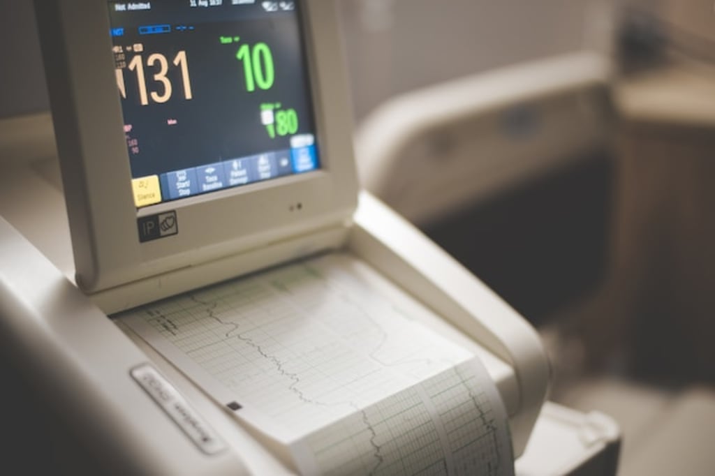
Ultrasound Machines: A Breakdown of Features and Applications
Exploring the Versatility of Ultrasound Machines: Features, Applications, and More

Ultrasound technology has revolutionized the medical field, providing invaluable diagnostic capabilities. From examining internal organs to monitoring pregnancies, ultrasound machines play a crucial role in healthcare. In this comprehensive guide, we will delve into the various features and applications of ultrasound machines, shedding light on their immense importance in modern medicine.
What is an Ultrasound Machine?
An ultrasound machine is a medical imaging device that utilizes sound waves to create real-time images of internal body structures. It works on the principle of sound wave reflection and echoes to generate visual representations of organs, tissues, and blood flow. These images, known as sonograms or ultrasounds, aid healthcare professionals in diagnosing and monitoring various medical conditions.
How Does an Ultrasound Machine Work?
Ultrasound machines consist of a transducer, a computer system, and a display screen. The transducer emits high-frequency sound waves into the body, which then bounce back as echoes when they encounter different tissues. These echoes are captured by the transducer and converted into electrical signals. The computer processes these signals and generates images on the display screen, allowing medical professionals to visualize the internal structures and identify abnormalities.
How to Choose the Right Ultrasound Machine
When choosing an ultrasound machine, it is crucial to consider your specific requirements, image quality, available features, transducer options, Doppler capabilities, portability, user interface, service, and support, as well as your budget. Understanding your primary usage and the medical specialty you will be focusing on will help narrow down the options. Prioritize high-resolution imaging and advanced features for accurate diagnosis, and evaluate the variety of transducers offered. Consider Doppler capabilities for vascular or cardiac imaging and assess the machine's portability and mobility based on your practice setting. A user-friendly interface and comprehensive service and support are also important factors to ensure a smooth experience. Finally, establish a budget and find a balance between cost and necessary features to make an informed decision.
Key Features of Ultrasound Machines
a. Transducer Types and Frequencies:
Ultrasound machines come with different types of transducers, including linear, convex, and phased array. Each transducer is designed for specific applications and offers unique advantages. Additionally, the transducers operate at different frequencies, ranging from 2 MHz to 18 MHz. Higher frequencies provide clearer images but have limited penetration, while lower frequencies offer greater depth but may sacrifice image resolution.
b. Imaging Modes:
Modern ultrasound machines offer several imaging modes to cater to diverse diagnostic needs. The two primary modes are 2D (two-dimensional) and Doppler. 2D mode displays cross-sectional images, providing a comprehensive view of organs and tissues. Doppler mode, on the other hand, assesses blood flow, helping identify abnormalities in vessels and organs like the heart.
c. Image Enhancement Techniques:
To improve image quality and accuracy, ultrasound machines employ various enhancement techniques. These include harmonic imaging, speckle reduction, and compound imaging. Harmonic imaging enhances image clarity by reducing artifacts, while speckle reduction minimizes noise interference. Compound imaging combines multiple angles to create a more detailed and comprehensive image.
Applications of Ultrasound Machines
a. Obstetrics and Gynecology:
Ultrasound plays a vital role in monitoring pregnancies and assessing fetal development. It enables visualization of the fetus, identification of abnormalities, and determination of the baby's position. Additionally, ultrasound assists in diagnosing gynecological conditions such as ovarian cysts, uterine fibroids, and endometriosis.
b. Cardiology:
Ultrasound machines are extensively used in cardiology to evaluate the structure and function of the heart. Echocardiography, a specialized ultrasound technique, allows physicians to assess heart chambers, valves, and blood flow. It aids in diagnosing heart conditions, including cardiac abnormalities, valve defects, and congestive heart failure.
c. Abdominal Imaging:
Ultrasound imaging of the abdomen helps in diagnosing conditions affecting the liver, gallbladder, pancreas, kidneys, and other organs. It enables the detection of liver cirrhosis, gallstones, kidney stones, and abdominal tumors. Moreover, ultrasound-guided procedures such as biopsies and drainages can be performed to obtain tissue samples or remove fluid accumulations.
d. Musculoskeletal System:
Ultrasound machines are valuable tools in evaluating musculoskeletal conditions, including sprains, strains, and joint inflammations. They aid in assessing soft tissues, muscles, tendons, ligaments, and joints. Ultrasound-guided injections and aspirations can be performed to deliver medication or extract fluid from affected areas with precision. This non-invasive and real-time imaging technique allows physicians to visualize the target area and ensure accurate needle placement.
e. Vascular Studies:
Ultrasound machines play a crucial role in assessing blood vessels and diagnosing vascular conditions. Doppler ultrasound, specifically, helps evaluate blood flow, identify blockages, and detect abnormalities such as blood clots or arterial stenosis. Vascular ultrasound is commonly used to examine carotid arteries, deep veins in the legs, and abdominal blood vessels.
f. Breast Imaging:
Ultrasound machines are often employed in conjunction with mammography for breast imaging. They help in differentiating between solid masses and cysts, guiding biopsies, and monitoring breast health. Breast ultrasound is particularly useful for younger women or women with dense breast tissue, as it provides additional information beyond mammography.
g. Emergency Medicine:
Ultrasound machines have become indispensable in emergency medicine, allowing rapid assessment and triage of patients. They aid in diagnosing conditions such as abdominal trauma, internal bleeding, or organ damage. Ultrasound imaging assists emergency physicians in making timely and accurate decisions, leading to improved patient outcomes.
Advancements in Ultrasound Technology
In recent years, ultrasound technology has witnessed significant advancements, enhancing its diagnostic capabilities further. Some notable advancements include:
a. 3D and 4D Imaging:
Traditional 2D ultrasound has evolved to include 3D and 4D imaging, providing more realistic and detailed representations of the examined structures. 3D imaging adds depth to the images, while 4D imaging introduces the time element, enabling real-time visualization of moving organs or the developing fetus.
b. Contrast-Enhanced Ultrasound (CEUS):
CEUS involves injecting a contrast agent into the patient's bloodstream to enhance the visibility of blood vessels and improve the characterization of lesions. It is particularly useful in liver imaging, aiding in the detection of liver tumors and monitoring treatment response.
c. Elastography
Elastography is a technique that measures tissue stiffness, providing valuable information about the elasticity of organs or masses. It helps in differentiating between benign and malignant lesions and assists in liver fibrosis assessment.
d. Portable and Handheld Ultrasound Devices:
The development of portable and handheld ultrasound devices has made ultrasound more accessible and convenient. These devices are lightweight, compact, and easily maneuverable, making them ideal for point-of-care diagnostics, emergency medicine, and remote or resource-limited settings.
Ultrasound machines have become indispensable tools in the field of medicine, enabling non-invasive and real-time imaging of internal structures. Their versatility spans across various medical specialties, from obstetrics and gynecology to cardiology, abdominal imaging, and musculoskeletal evaluations. With advancements in technology, including 3D/4D imaging, contrast-enhanced ultrasound, and elastography, ultrasound machines continue to evolve, providing enhanced diagnostic capabilities. As a cornerstone of modern medical practice, ultrasound machines empower healthcare professionals to make accurate diagnoses, guide interventions, and improve patient care.


Comments
There are no comments for this story
Be the first to respond and start the conversation.