
Soft tissues and Neuro-vascular structures
Neuro-vascular structures

A comprehensive understanding of structures at risk is essential for the treatment of pelvic fractures. Because the large number and the high density of para-pelvic organs and soft tissues not only play a vital role in the integrity of ligaments to pelvic but also has its significant role in relation to the acute prognosis for e.g. hemorrhage and late prognosis for e.g. urological and neurological, injuries.
The rate of mortality has been increased due to complex pelvic injuries such as: combination of osteoligamentous and concomitant para-pelvic soft-tissue injuries like urogenital, hollow-visceral, and neurovascular. And if an unstable hemodynamics is caused by life-threatening hemorrhage, then mortality rate is higher. That’s why, special consideration must be given to the patients who have been admitted with a blood loss of more than 2,000 ml.
Terminology and Classification
The first and foremost thing is identification of the grade of stability or instability of the fracture. Clinical and radiological assessment of the pelvic is greatly dependent on it. Three different degrees of stability or instability which are the basis of indications are as follows:
Mechanical structure of the pelvic ring intact: Type A injury (incidence 50% –70% of patients).
Partial posterior stability, rotational instability: Type B injuries (incidence 20% –30% of patients).
Combined anterior and posterior instability, translational instability: Type C (incidence 10–20% of patients).
The type of fracture is the determinant factor of the treatment:
Type A injuries: Surgical stabilization is exceptionally suggested in these fractures.
Type B injuries: Stabilization of the anterior pelvic ring alone is enough in such cases.
Type C injuries: Adequate stabilization of the ring is required in these fractures so that the risk of secondary displacement can be minimized. However, differentiation of partial or complete posterior instability (type B or C) may be abstruse.
The primary assessment and evaluation should be done carefully and rechecked. Review of diagnosis and classification can be done if required and then the therapeutic decisions must be taken. In the last 30 years, more than 40 classification systems or important modifications have been documented.
As compared to other body regions, pelvic injury may have variations such as: multiple lesions of the anterior and/or posterior ring, of the right and/or left side, and various fracture patterns and anatomical segments. The present classification of pelvic injuries is based on AO Muller system which is derived from the assessment of the injury mechanism and stability or instability of the pelvic ring occurred due to it. The three basic fracture types A, B, C are further categorized in groups, sub-groups, and specific modifiers. Thus, classification of each injury and combination of injury is done in alphanumeric symbols.
Primary evaluation and decision making
The assessment of pelvic injuries at primary level helps to know whether severe internal hemorrhage is the resultant of pelvic fracture or not. It also helps to assess the degree of mechanical stability of the pelvic ring clinically and radiologically as well. The diagnosis para-pelvic soft-tissue and organ injuries is also done at this level.
Immediate surgical resuscitation procedures ideally by following a protocol or algorithm must be done if hemodynamic instability of pelvic is diagnosed. In most of the cases of pelvic injury, hemodynamics is affected either minimally or not at all. Although a detailed clinical and radiological work-up is required for diagnosing and grading of a pelvic injury.
Clinical examination
AP and lateral compression can be used for the manual examination of pelvic stability. Occasional push-pull examination of the leg can be done additionally. But in case of unstable patient, to prevent further induction of blood loss, repeated examinations should not be done.
Radiological examination
For the radiological examination, the AP pelvic view is required it can provide a reliable working diagnosis in about 90% of the cases. Oblique views are required to assess anterior, posterior, rotational and craniocaudal displacement for three-dimensional analysis.
In all cases, a CT examination is required to precise the posterior pelvic injury and associated acetabular fracture if any. CT is not used for immediate assessment so if required, it can be delayed until the stabilization of the patient.
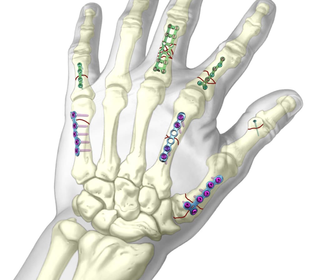
Orthopedic implants suppliers in UK are providing wide range of trauma implants and instruments such as: Radial head prosthesis, Microlock Locking Hand Implants, LCP, screws and nails which are used for different type of fractures
About the Creator
Siora Surgicals
Siora Surgicals is an Orthopedic Implants Manufacturers and Suppliers , selling trauma Implants and Instruments in India. We are one of the best manufacturing Orthopedic Implants Instruments as per quality management system.
Keep reading
More stories from writers in Longevity and other communities.
Pints & Parkruns: Jubilee, Spennymoor
If MC Escher created a parkrun, it might look a bit like Jubilee. Based in a compact – but surprisingly lovely – park in the small County Durham town of Spennymoor, it twists and turns its way up repeated hills. Although basic physics says it must come down again, somehow this route never feels like it gives runners a proper descent.
By Andy Potts6 days ago in Longevity
Diabolical Dealing
Charlene Lind found herself at the home of her therapist, Dr. Alexandra Vech, who she had been seeing for a week. What was supposed to be a session ended up becoming a desperate plea of help from Charlene as she was suddenly losing consciousness. She was cognizant enough to know that she had only a minute left until she would be out like a light.
By Clyde E. Dawkins7 days ago in Fiction


Comments
There are no comments for this story
Be the first to respond and start the conversation.