
Is It Possible To Detect Fatal Growth Abnormalities Through CT Scans?
- A Must Read

Advancements in medical imaging have revolutionized the field of prenatal diagnosis, enabling healthcare professionals to monitor foetal development and detect potential abnormalities.
Among the various imaging techniques available, computed tomography (CT) scans have gained attention for their ability to provide detailed cross-sectional images of the body.
However, when it comes to detecting foetal growth abnormalities, the role of CT scans is relatively limited.
This article aims to explore the possibilities and limitations of using CT scans for identifying foetal growth abnormalities.
Understanding CT Scans
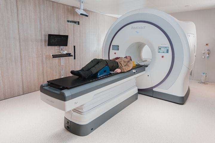
Computed tomography, commonly known as CT, utilizes a series of X-ray images taken from different angles to create detailed cross-sectional images of the body.
These images are then reconstructed using computer algorithms, allowing healthcare professionals to visualize the internal structures with greater precision.
CT scans are widely used in diagnosing various conditions, such as detecting tutors, assessing bone fractures, and examining the organs.
Challenges With foetal CT Scans
While CT scans have proven to be highly effective in many areas of medical diagnosis, their use in foetal imaging is limited due to several factors.
First and foremost, CT scans involve exposing the foetus to ionizing radiation, which carries potential risks, particularly during crucial stages of development.
Radiation exposure may increase the risk of cancer or have adverse effects on the developing foetus.
Consequently, CT scans are generally avoided unless there is a compelling medical indication that outweighs the potential risks.
Alternative Techniques For Foetal Imaging
To address the concerns associated with radiation exposure, alternative imaging techniques are preferred for foetal evaluation.
Ultrasound remains the primary imaging modality for monitoring foetal growth and detecting abnormalities.
Ultrasound is non-invasive, does not involve ionizing radiation, and provides valuable information regarding foetal development, including measurements of foetal size, organ formation, and overall well-being.
Other specialized imaging techniques, such as magnetic resonance imaging (MRI), have also gained popularity in foetal imaging.
MRI offers detailed images of the developing foetus, enabling healthcare professionals to assess foetal growth, identify structural anomalies, and evaluate organ development.
Unlike CT scans, MRI does not involve radiation exposure, making it a safer alternative for foetal imaging.
The Role Of CT Scans In Specific Cases
Although CT scans are not routinely used for foetal imaging, there are specific scenarios where they may be considered.
In rare cases, when there is a suspected abnormality that cannot be adequately visualized through ultrasound or MRI, a CT scan may be used as a last resort.
This is typically done when the potential benefits outweigh the risks and when the information obtained is critical for determining appropriate medical management.
For example, in cases of suspected skeletal abnormalities, where bone structures are difficult to assess using ultrasound, CT scans may be utilized to obtain more detailed information.
Similarly, in cases of complex congenital heart defects, CT scans can provide additional insights into the heart's structures and help guide surgical interventions.
Conclusion
In conclusion, while computed tomography (CT) scans offer exceptional imaging capabilities in various medical fields, their application in foetal imaging and detecting growth abnormalities is limited.
The potential risks associated with radiation exposure make CT scans less favorable compared to other imaging techniques, such as ultrasound and magnetic resonance imaging (MRI).
Ultrasound remains the primary choice for monitoring foetal growth and detecting abnormalities, while MRI provides detailed information without radiation exposure.
It is essential for healthcare professionals to carefully weigh the benefits and risks before considering a CT scan for foetal evaluation.
The decision to use a CT scan should only be made when there are specific medical indications that cannot be adequately addressed by alternative imaging methods.
As technology advances, it is likely that safer and more specialized imaging techniques will continue to emerge, further enhancing our ability to detect and manage foetal growth abnormalities with minimal risks to both mother and child.
Are you expecting? Are you seeking a reliable diagnostic center to undergo a CT scan? If you're located in Australia, look no further. Click on this link to access top-notch diagnostic imaging technology and expert professionals. And that's not all! They have multiple centers conveniently located throughout the country.
About the Creator
Christopher David
I am a writer, editor and an avid reader.
Keep reading
More stories from Christopher David and writers in Longevity and other communities.
Can You Drink Wine If You Have Diabetes?
Whether you're hanging with your friends or enjoying a movie night with your partner, a popular way to unwind is to sip a glass of wine. Yet if you have diabetes, you're probably wondering whether you can enjoy an after-dinner glass, too. After all, you might assume that wine is loaded with sugar, making it taboo in your diet.
By Kaly Johnes7 days ago in Longevity
Final Call
She flings her arms around my neck and squeezes me like she’s never going to let me go. My head spins as her lips brush against my cheek. She must be able to feel my heart thumping in my chest. I shuffle backwards and ram my hands into my coat pockets, watching her reach down and heave the rucksack across her shoulders.
By Caroline Cravena day ago in Fiction


Comments
There are no comments for this story
Be the first to respond and start the conversation.