
The Robin Hood of diseases
How a disease of the brain can save your arm.

The human body is a fascinating subject. Here's just one example testifying to the statement.
Before, we dive into the disease itself, I want to do a little recap of the normal functioning of the body, to put the disease in perspective later.

This is a MRA (Magnetic Resonance Angiogram) showing the vasculature of the normal human body. The bright lines of varying thickness are blood vessels filled with blood of corresponding thickness. Notice how brightly the brain is lit up compared to the rest of the body (save for the heart and the great vessels). Despite making up only 2% of the body weight, the brain consumes nearly 15 to 20% of the cardiac output (volume of blood pumped out of the left ventricle of the heart in a minute). Without it, the brain cells start dying.
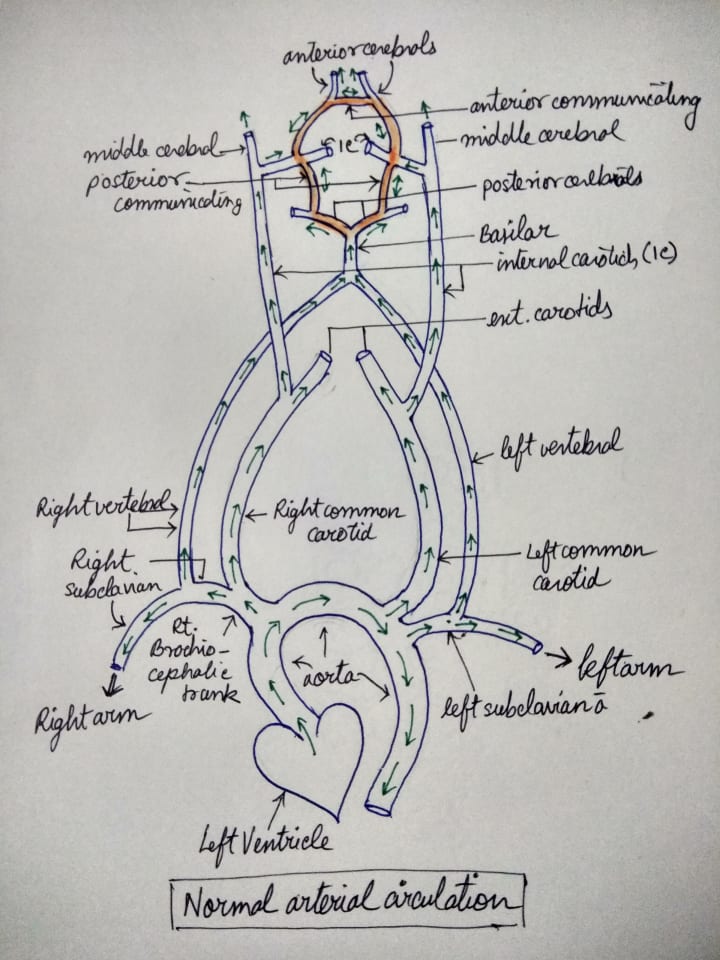
This is a quick sketch I made for this answer. Excuse the poor handwriting— I am a medical student, I get a free pass. This highly schematic diagram represents the first few major branches of the aorta before it descends, (i.e, the right brachio-cephalic trunk, the left subclavian and the left common carotid arteries) and an empirical blood supply of the brain. The right brachio-cephalic trunk divides into the right subclavian and the common carotid arteries. The carotids, as is common knowledge, supplies blood to the neck and head. The subclavian gives off a few branches before becoming the brachial artery— the principal artery to the upper limbs. One of the branches of the subclavian is the vertebral artery, which again supplies the brain through the basilar artery. Green arrows mark the direction of flow of blood.
Now, what I have marked in the above image in a slight shade of orange is the Circle of Willis. This is a network of circulatory anastomosis that supplies the different parts of the brain and its surrounding structures. The image below is testament to how complex the cerebral vasculature actually is. Note the basilar artery and its branches.

With all this in mind, imagine this scenario:
You are at the gym, doing some bicep curls. You are lifting more weights than usual to impress that cute girl on the elliptical. (That rhymes, holy shit!) Out of the blue, you feel a sharp pain shooting down your left arm. You think you pulled a muscle, but before you can put the weights down, it’s you on the ground on your knees- hurling your stomach contents out on the gym floor. You have no idea about why in hell is the world spinning so fast and barely manage to blurt out your symptoms to the gym trainer before passing out in the pool of your own puke.
So much for impressing the girl.
Fearing a heart attack, the gym trainer immediately calls for an ambulance. You are in a hospital within 45 minutes, but much to the surprise of the ER doctor and you, your ECG is normal and markers for acute MI are negative. The doc orders a Doppler study of your blood vessels and it turns out that the root of your left subclavian is blocked by a stenosis/atheroma.
So why in the world did decreased blood supply to your arm cause such explosive neurological symptoms like extreme dizziness and nausea?
This, is the answer.
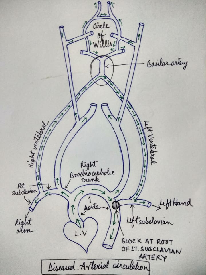
Whenever the root of the subclavian artery is blocked or stenosed, the blood pressure beyond the block falls drastically. This severe fall in BP creates a pressure gradient from the high pressure basilar artery to the now low pressure subclavian artery, so much so, that the direction of blood flow reverses in the ipsilateral vertebral artery— “stealing” blood from the Circle of Willis via the basilar artery and redirecting it to the subclavian artery beyond the block. Given these properties of this disease, it has been aptly named “Subclavian Steal Syndrome”.
The basilar artery supplies oxygen rich blood to the brainstem and cerebellum. Since redirecting blood deprives this artery of its quota of blood, the hypoxia to the brainstem and cerebellum results in the aforementioned neurological symptoms. But your mental faculty remains intact because the forebrain/cerebrum is supplied by branches of the internal carotid artery, which are absolutely A-OK.

What fascinates me most about this disease is the fact that this disease chucks the notion of “blood in vessels always flows in a unidirectional manner” right out of the window. Given half the chance, these sneaky bastards would do opposite of what they are supposed to be doing— in some cases, saving a limb from atrophy, paralysis or in a worst case scenario, gangrene.
About the Creator
Diptangshu Karmakar
Medical student. Certified nincompoop. Questionable bookworm and painter. Common sense: defenestrated. Interested in anthropology, evolution and history, especially military.
Keep reading
More stories from Diptangshu Karmakar and writers in Longevity and other communities.
Shades of Skin
As a freshman high school student, I remember this question popping up during the physics class on radiation: When black catches heat faster and leaves faster (opposite to white),why didn't people who live in hot areas and especially work under sun didn't evolve to have lighter skin to avoid sun strokes, and it only becomes only darker under the sun?
By Diptangshu Karmakar4 years ago in Futurism
Overlooked Urine at Night: A Vital Window into Your Health
Daniel, aged 65, has consistently maintained a habit of exercising, resulting in a much more robust physique compared to his peers. Approximately six months ago, he began experiencing symptoms of hematuria during urination. Initially, he dismissed it as insignificant. However, as time passed, the frequency of hematuria increased, and later stages even saw the passage of blood clots. It was then that Daniel realized something was amiss and sought medical attention.
By Amanda Chou4 days ago in Longevity
Reaching Out
I promise her. I'd do anything for her. She's my mom. Even as Lanie and Deanna are flying home, Mom is scrappy fighting dying. She lays too still in that too-big bed with all the toasty white hospital blankets, in the south tower, at the broad end of a long slow-turning corner that delivers me again to her private room with the view she can't see through, with the beeping that tells us nothing new, and all these ice chips she can't swallow, and a flood of well-intentioned nurses who cannot do a damned thing all the same.
By Christy Munsona day ago in Fiction


Comments
There are no comments for this story
Be the first to respond and start the conversation.