
Structure Of Human Eye with well labelled diagram
Human Eye

Humans have binocular vision. The eye can segregate tone, evaluate length, width and profundity outwardly, and structure a genuine altered picture.
The eyes are two in number and held up in circles (hard attachment) of the skull. The eye is an empty, round organ, around 2.5 cm in breadth and around 6 to 8 grams in weight. It has two sections -
(1) Protective gadgets: The eye has four defensive gadgets.
(I) Eye temples: The apparently coordinated hair of the eyebrows convey the perspiration and downpour drops streaming down the brow to the sides to forestall their falling into the eyes.
(ii) Eye tops (Palpebrae): In man two eyelids are available, the upper is portable. They are routinely shut at short stretches to clean the cornea. This is called squinting. In frogs two upper eyelids are unflinching and the lower eyelid is versatile. The nictitating layer is available in frogs which safeguard the eye in the water. Development of the nictitating layer happens by retractor bulbi. It becomes collapsed by levator bulbs.
A nonfunctional vestigial nictitating layer, called plica semilunaris, happens in natural eyes. It remains forever withdrawn at the internal point of each eye.
(iii) Eyelashes: The eyelids bear at the free edge a column of solid hair, the eyelashes. These check the section of residue particles, small bugs, and downpour drop into the eyes.
(iv) Eye organs
(a) Meibomian organ: The eye-covers bear at the free edge a line of the meibomian organ that is changed sebaceous organ. (Go about as a lubricant).
(b) Lacrimal organ or Tear organ: It lies in the upper external piece of the circle and secretes a marginally saline, watery liquid that contains a bacteriolytic catalyst named lysozyme. This discharge saturates the outer layer of the eyeball. The overabundance of this discharge goes through the nasolacrimal channel. It is a changed perspiration organ.
(c) Harderian organ: Some amphibian well evolved creatures (whale) have harderian organs which grease up the nictitating film. It is additionally found in frogs and birds.
(d) Glands of Zeis (zis): These are adjusted sebaceous organs, found at the base of the hair follicle of eyelashes, pour greasing up liquid in the hair follicle. The disease of these organs is a pen.
(e) Glands of Moll: It is an adjusted perspiration organ and opens into the follicles of eyelashes.
In human meibomian, lachrymal, Moll's organs, and zeis organs are available.
(v) Connective tissue: A layer of greasy connective tissue encompasses the eyeball. It fills in as a delicate shockproof cushion.
(2) Eyeball: Eyeball is comprised of 3 coats or tunic.
(I) Sclerotic layer (Fibrous tunica): Outermost and dark, sinewy, and non-vascular layer effectively considered white of the eye. It is a layer of thick connective tissue comprised of collagen filaments and fibroblasts. Sclera covers the whole eyeball aside from the cornea and gives shape to the eyeball. Sclera in frogs is cartilaginous.
(a) Cornea: In the middle, the sclerotic layer converges with the straightforward round window called the cornea.
(b) Conjunctiva: The cornea and uncovered piece of the sclera is covered remotely by a slender, straightforward layer of the conjunctiva.
(ii) Choroid layer (Vascular tunica): Also known as uvea center. It is a vascular layer that supplies supplements to the eye. It is recognized in three sections choroid, ciliary body, and iris.
(b) Choroid: It is an exceptionally vascular back piece of the vascular tunic. The choroid happens in the principle of a piece of eyeball stuck to the sclerotic. (The shade is rosy in hare and dark, brown, or somewhat blue in man).
(b) Ciliary body: Ciliary body is vascular and pigmented like a choroid, comprised of ciliary cycles and ciliary muscles (just roundabout sort). The ciliary body is concealed by the iris. The ciliary body helps inconvenience by adjusting the focal point of the eye from an object or the state of focal point close or far vision.
(c) Iris: Beyond the ciliary body, the vascular tunic strongly turns inwards, shaping around, a rack-like stomach called the iris. The shade of the iris is answerable for the shade of the eye e.g., brown, dark, blue, or green. In pale-skinned people, the iris is insufficient shades.
Focal point: Lens is a dreary, straightforward, and sinewy crystalline structure comprised of protein (an and b glasslike protein) and encased in a focal point layer. It is ectodermal in beginning. The focal point is held up in the eyeball by the suspensory tendon of the ciliary body. Suspensory tendons are known as "Zonula of Zinn". In man focal point is biconvex while in the frog it is curved (subspherical).
Focal point partitions the eyeball into 2 chambers external fluid chamber (somewhat isolated into an enormous front and a more modest back chamber) loaded up with watery humor (watery) framed by the ciliary body and an inward glassy chamber loaded up with glassy jam (or Wharton's jam) containing close to 100% water, some salt a little mucoprotein (vitrein) and hyaluronic acid
Differences between Aqueous Humour and Vitreous Humour Read More
About the Creator
Keep reading
More stories from NEET RANGERS and writers in Longevity and other communities.
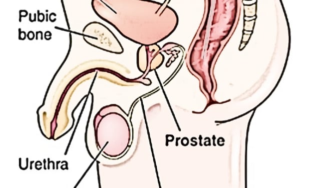
What is Testes and cryptorchid and testis structure
The testicles, otherwise called balls or male balls, lie behind the penis in a pocket of skin called the scrotum. The testicles move openly in the scrotum yet every testis is connected to the body divider by a slender string called the spermatic rope, which goes through a cavity in the pelvis and into the midsection. The line contains the nerves and veins for the testicles along with the vas deferens, which convey the sperm from the testicles into the urethra; the urethra is the way for sperm to the beyond the body at discharge.
By NEET RANGERS2 years ago in Longevity
The Health Benefits of Cinnamon
What is Cinnamon? According to Wikipedia, cinnamon is a spice obtained from the inner bark of several tree species from the genus Cinnamomum. It is the name for several species of trees and the commercial spice products that some of them produce (en.wikipedia.org/wiki/cinnamon).
By Lisa Briskey2 days ago in Longevity


Comments
There are no comments for this story
Be the first to respond and start the conversation.