
Silk & Diamonds: The Fancy End of High-Tech Medicine
It turns out silk, an ancient natural fibre, has versatile uses in medicine — especially when combined with diamonds.

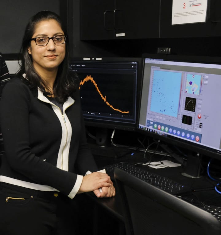
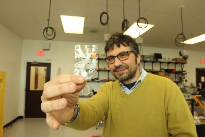
ASMA KHALID enjoys wearing a silk dress, and appreciates diamonds for their beauty. But she never expected both would end up being the cornerstone of her work as a physicist. Yet they have, and opened up a whole new way to see deep in the body and even deliver drugs.
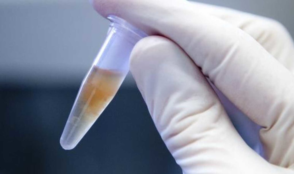
As a PhD student at the University of Melbourne in Australia, she had been working with nano-diamonds, particles of solid carbon arranged in a crystal structure that are less than one thousandth of a millimetre in size. Because they’re inert to organic processes and and have excellent light-emitting properties, nano-diamonds are being widely explored in biology as sensitive tools for diagnostic imaging and sensing.
“Nano-diamonds are fluorescent, they glow brightly when we excite them with a laser. But they have a rough surface and tend to clump together a lot, sometimes blinking when we want them to fluoresce,” said Khalid, now a postdoctoral fellow at Melbourne’s RMIT University and an associate investigator at the Centre for Nanoscale BioPhotonics (CNBP).
“We wanted to improve their surface and optical properties by coating them with a material that was still biocompatible in the body. That’s when we tried silk.”

Silk as a fabric dates back 12,000 years; it was first used by the Yangshao, a Neolithic people living around the Yellow River in China. The best-known silk is extracted from the larval cocoons of the mulberry silkworm, which is reared in captivity. Its shimmering appearance is due to the prism-like structure of silk fibre, which allows it to refract incoming light at different angles, producing a range of colours.
As a fibre, it is not harmful or toxic to living tissue because it’s made entirely of proteins and amino acids — and it also has great optical properties, added Khalid. “Silk actually enhances the brightness of the nano-diamonds significantly. And when we implanted a silk-coated hybrid inside mice, we found the silk dissolved in the body without causing any inflammation.”
Her resulting paper in Biomedical Optics Express generated a lot of interest, leading Khalid to a scholarship at Prof Fiorenzo Omenetto’s Silk Lab at Tufts University in Boston, which has pioneered the use of silk in photonics and biotechnology. There, she leaned to make optically transparent silk, which worked even better as biocompatible cladding.
“I learned how to extract silk from cocoons and transform that liquid silk into a range of different structures, like implantable films, injectable nanoparticles, 3D printed frames, and several other structures and devices for biophotonics and biomedical applications,” she recounted.
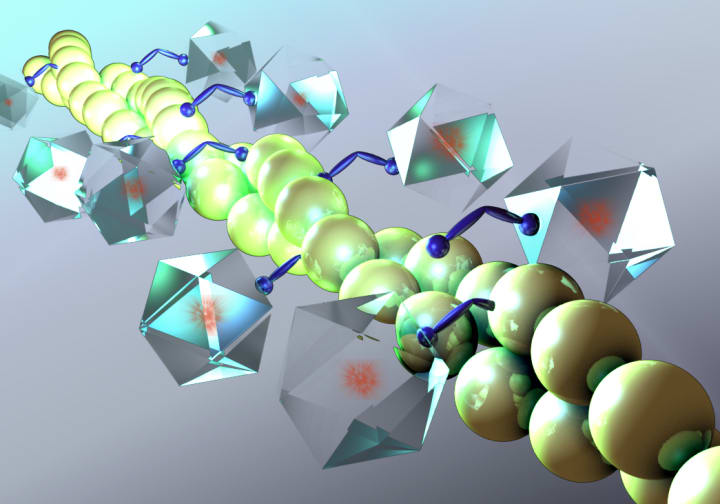
“I also produced silk-coated nano-diamond spheres, which worked really well as super-bright cell imaging tools, and drug-loaded nano-diamond silk spheres that could be used as vehicles for controlled release of drugs in anticancer treatment,” she said.
“The hybrid spheres can release small amounts of drug over the period of weeks as the silk dissolves, and because the nano-diamonds fluoresce, we can track the release of the drug.”
The work led to two new scientific papers, and also piqued the interest of Tufts University in the optical properties of nano-diamonds as imaging and biosensing tools. When Khalid returned to Australia and joined RMIT, she brought the Tufts collaboration with her. “So, we combined our work and got very interesting applications in imaging, sensing, drug release and tissue regeneration.”
Khalid started her own lab at CNBP’s RMIT node to produce liquid silk nanoparticles from cocoons, creating fibre coatings, and electro-spun membranes — sheets of interconnected, uniformly sized pores that can be combined with a host of useful particles for health monitoring applications.
At the time, Dr Jiawen Li, a biomedical engineer at the University of Adelaide’s CNBP node, was experimenting with a new class of tiny fibres inside ultra-thin fibre endoscopes that could take measurements deep in the body of a living organism, rather than from excised samples.
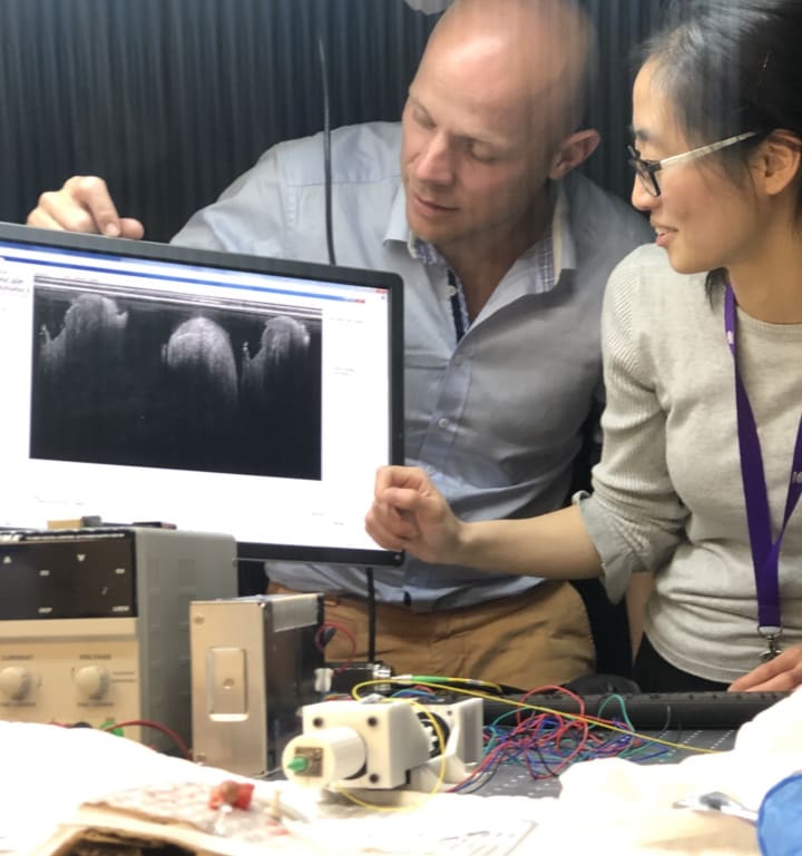
“It might be a tumour deep in the lung and we want to know whether it’s malignant or not by measuring pH level, oxygen, carbon dioxide or other target molecules, but without having to remove a piece of tissue for biopsy,” she said.
“Or instead of drawing blood and then getting the chemical composition of something like calcium or glucose, we want to insert these really tiny optical fibres, smaller than a human hair, which are minimally invasive, to take real-time localised measurements at its original position without the need to wait for the lab result.”
The fibres had to be sheathed so they didn’t chemically interact with living tissue, so Li had been exploring polymers, synthetic organic compounds with molecules bonded together in long chains and used as plastics and resins.
“The problem is that to enable these fibres to take measurements, you usually need to use harsh chemicals, which is not good for putting into the body,” Li added. “Or those chemicals might interfere with the information we’re trying to collect, so that you’re changing what you’re trying to observe.”
Khalid suggested using silk, and the duo tried coating Li’s optical fibres with silk and testing them for biosensing and bioimaging in mice. “I couldn’t believe how simple it was,” Li said. “The silk is totally biocompatible; it’s not treated as a foreign intruder. And the whole process of adding sensors, like fluorophores, on top of the fibre is done at room temperature by just dipping the fibre into solution for 30 seconds.”
The next challenge was ensuring that when incorporating biomarkers into silk, the sensors stayed attached and worked as expected. That’s when organic chemists Aimee Horsfall and Patrick Capon, PhD students in the CNBP’s biosensors group at the University of Adelaide, joined the effort.
“We needed to test the robustness of the system,” said Horsfall. “If we poke it into somebody to reach an organ or tumour, will the biomarkers stay attached and function properly, and what are the limitations of the system?”
They started with a sensor for pH, a chemical scale used to specify how acidic or alkaline a water-based solution is. But making the silk cladding work as a reliable sensor housing turned out to be a challenge: no matter how many ways they tweaked the chemistry of the silk, not all of the biomarkers stuck fast. After months of dead ends, they changed tack, and looked for a bonding peptide that might help anchor the sensors directly to the silk.
After bathing the fibre in a mixture of the silk and sensors in silk-binding peptide for 30 seconds, the critical step turned out to be immediately dipping the silk-coated fibres into methanol for another 10 seconds.
“That changes the structure of the silk and makes it much more crystalline, which really attaches it to the fibre strongly,” said Capon. “We’ve tested the fibre and verified we can hook up a laser and get a fluorescent signal from the sensor that’s anchored there. And we’ve found the only way we’re able to make the bonding fail is to literally break the fibre.”
Now that the researchers have proven sensors can be dependably attached, they’re expanding from pH to hydrogen peroxide sensors for sperm cell health; sensors for metal ions like calcium and zinc for use in fertility and in vitro fertilisation; in fact, a whole range of applications to target specific ailments and conditions. Clearly, silk is going to be the workhorse for sensor work in fibre optics.
“There’s a lot of work to do, but we’re pretty excited,” said Horsfall. Capon agreed: “Every time we talk, we seem to come up with another three new ideas. How many sensors can we pack onto a fibre? What kind of applications can we find that biologists are interested in, or that clinical staff need?”
Meanwhile, Khalid has since expanded her work into silk-coated fluorescent nanoparticles for cancer imaging in collaboration with CNBP’s Macquarie University node in Sydney; silk-coated magnetic particles for brain imaging with an engineering group at RMIT; and with the University of Melbourne collaboration to create metal oxide-silk nanoparticles for cell imaging.
She is also developing other exciting hybrid silk materials to address the current limitations in wound and burn care technologies.
“I always used to think of silk as a fabric,” Khalid mused. “But when I did the literature review, I realised that it’s been used in medicine for centuries as suture. Silk and diamond may be fancy materials, but they also have great optical properties and a lot of benefits in health and medicine.”
Like this story? Please click the ♥︎ below, or send me a tip. And thanks 😊
About the Creator
Wilson da Silva
Wilson da Silva is a science journalist in Sydney | www.wilsondasilva.com | https://bit.ly/3kIF1SO
Keep reading
More stories from Wilson da Silva and writers in Longevity and other communities.
Making Nanomachines That Cure from the Inside
THE AMBITION is grand and the timeline spans decades: the development of nanomachines dispersed in the human body, performing detection, diagnosis and treatment and communicating wirelessly with physicians who monitor and direct treatment.
By Wilson da Silva3 years ago in Longevity
The Curious Case of My Hair Growth Gummies Adventure
Chapter 1: The Great Hair Crisis It all began on a gloomy Tuesday morning. I stood in front of the bathroom mirror, my damp hair clinging to my forehead like a clingy ex. The reflection staring back at me was a sad version of my former self. My once-voluminous locks had decided to play hide-and-seek, and they were winning. The widening part mocked me, and the receding hairline whispered secrets I didn’t want to hear.
By health_kkkkeep3 days ago in Longevity


Comments
There are no comments for this story
Be the first to respond and start the conversation.