


Every year, when children are on summer vacation, it is also the busiest time for dermatology clinics. There are two main types of common skin problems, one is mosquito bites, unknowingly "planted strawberries" on the body, and red and itchy bulges. The second is the removal of spots and moles. Many young people are intolerant of the small black spots on their faces and bodies and want to "get rid of them before they go".
These skin "small" problems, some hidden "big" danger.
What are the dangers of mosquito bites?
Commonly caused by allergic insects are mosquitoes, fleas, bedbugs, mites, cryptographers, etc. Mosquitoes, fleas, bedbugs, and mites have common features, are in the bite, their saliva and toxins into the human skin, stimulating the skin to produce local allergic reactions, usually in the bite site redness, ampules, blisters, large bubbles, redness, and swelling from the size of soybean to the adult palm, infants and children usually react more obviously, itching or burning pain is obvious, and even cause systemic allergic reactions. These types of bug bites have similar localized rashes, so only the discovery of the bug body can confirm the diagnosis of some type of bug bite dermatitis.
If only small redness, pa-pules, pa pules, can be applied externally arthritis, furnace glycol, hormone-containing ointment, such as dermatology, Fuji cream, momentariness floater, etc., generally 3 - 4 days after the gradual improvement, if the redness and swelling range is larger, multiple blisters or even large bubbles, especially infants and children caused by crying more than, to the hospital promptly, anti-allergy drugs can be given orally, local symptomatic treatment.
Some patients shoot the insects to death on the skin, and the limbs of the insects may remain within the skin. There was a patient, one month after the bite, external application of various ointments still can not effectively stop the itch, coin-sized lumps, surface scans retching, to our department, microscopy found a very short and thin brown thing in the center of the lesion, cotton swabs wiped away a week after the healing. Consider a persistent allergic reaction caused by insect limbs remaining within the skin.
Can small moles be removed as often as you want?
1. Causes and factors related to the development
Pigmented nevus is a developmental malformation, which is caused by the abnormal movement of melanomas from the neural crest to the epidermis, resulting in the local aggregation of melanomas, and its incidence is related to age, race, and genetic and environmental factors. Pigmented nevi can appear in early childhood, increase rapidly during adolescence, reach their maximum number at the age of 20-29, and then gradually fade with age. The incidence of pigmented nevi is higher in Caucasians, but pigmented nevi in the plantar, nail bed, and mucous areas are more common in Blacks and Asians. Pigmented nevi tend to be more prevalent in certain families, especially in familial melanoma. Environmental factors, such as sun exposure, can increase the number of pigmented nevi in exposed areas.
2. Pigmented nevus classification
Pigmented nevi are divided into junction nevi, mixed nevi, and Alderamin nevi according to the depth of their distribution on the skin.
Junction nevus is present at birth or occurs soon after birth, small, less than 6 mm in diameter, smooth and hairless, flat or slightly higher than the skin surface, light brown to dark brown, and can occur on any part of the body. Mixed nevus is slightly higher than the skin surface, and some of them have hair penetration, which is mostly seen in children and adolescents. Alderamin nevi are more common in adults, are hemispherical, and are lighter in color than the first two, or even skin-colored. Cross border nevi have a higher chance of malignant transformation and should be removed with caution by laser.
Causes of pigmented nevus formation and growth
(1) Sun exposure
Sun exposure causes multiple or severe sunburns, and some outdoor workers are prone to develop pigmented nevi due to long-term sun exposure. Exposure to sunlight in the middle of holidays, summer vacation outings, beach surfing, sunbathing, etc. Neonatal physiotherapy
(2) Skin damage
a. Various reasons for the formation of blister blisters. Skin damage and blisters are caused by strong acid, strong alkali, and toxic gas stimulation.
b. Skin damage caused by various skin diseases, toxic epidermal electrolysis, heretic epidermis bullock, and severe drug allergy.
c. Sclerosis strophic moss, scar formation.
(3) Systemic suppression
a. Chemotherapy, especially in children with malignant hematologic diseases
b. Solid organ transplantation (kidney transplantation), AIDS
(4) Elevated hormone levels
a. Pregnancy
b. Growth hormone, which promotes the increase in the size of pigmented nevi, but not the number
c. Thyroid hormone, hyperthyroidism
d. Some other diseases such as epilepsy or EEG abnormalities.
Are all small dark spots and patches on the skin surface pigmented nevi?
Pigmented nevi need to be distinguished from some diseases: junction nevi in childhood are distinguished from freckles and nigga, which increase slightly and gradually with age, while nigga usually remain unchanged. Mixed nevus and Alderamin nevus are distinguished from seborrhea keratitis, pigmented basal cell carcinoma, dermatologist, neuroticism, etc. Seborrhea keratitis has a rough appearance with wart-like growths; dermatologist is hard in texture with skin depressions and is commonly found on the lower extremity, and a normal-skinned or mildly pigmented intro-dermal nevus with a tip is difficult to differentiate from neuroticism.
How to distinguish pigmented nevus from other diseases by examination?
Electronic microscopy has been widely used for the diagnosis and differential diagnosis of skin diseases. Normally pigmented nevus microscopically: the pigment is uniformly reticulated, spherical, or pebble-shaped, with a light to dark brown color and some small thread-like blood vessels in the middle. If you see large dark pigmented masses with uniform black color or richer color with two or more colors such as gray-blue, blue-white, red, etc., and more large dendrite blood vessels or redder bases under the microscope, be highly alert.
Can pigmented nevus be removed by laser or freezing?
Pigmented nevi on the face affect the aesthetics, which is the main reason why many patients visit the clinic. The answer is, not all pigmented nevi can be removed by simple methods. They can be removed by laser only if they are less than 3 mm in diameter, have a uniform color, neat edges, smooth surface, soft epidermis, constant size and color, and appear under dermatology as uniformly distributed pigmented clusters with a single color and only a few small blood vessels, and generally do not need to be frozen. Before the laser, we will communicate with the patient to do a larger and deeper range, and try to remove it cleanly at one time. For recurrent pigmented nevi, patients are mobilized to remove them surgically as much as possible to avoid repeated stimulation by other means and increase the chance of malignant changes.
What changes in pigmented nevus need to be removed surgically?
When a pigmented nevus suddenly increases in size, deepens in color, has redness at the edge, has to crust on the surface, has itching or pain, and has small satellite loci around it, you should seek medical consultation and do a stereoscopic examination at the same time, and if there are abnormal changes under the mirror, it should be removed as soon as possible. Plantar, waist circumference, maxilla, groin, neck, shoulder, and other parts that are easily damaged by friction should be closely observed, and some with irregular edges, uneven color, and diameter greater than 6 mm should be paid more attention to and should be promptly ore-screened by dermatology at the hospital to determine whether to operate or not and should be promptly removed once it is found to be rapidly increasing, partially elevated or with ulceration and bleeding.
How should we take care of the skin every day in order not to stimulate pigmented nevus?
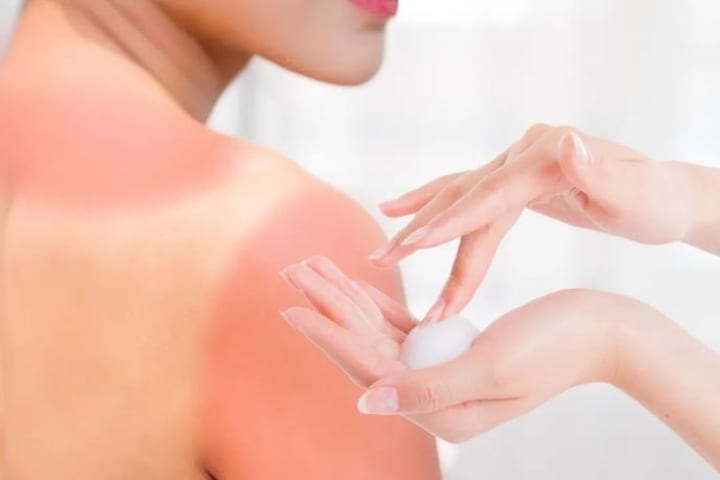
(1) Avoid sun exposure and apply sunscreen when you go out.
(2) Use mild cleansers and body washes, and do not often use irritating or alkaline ones.
(3) Choose skin care products with good moisturizing performance according to your skin, and remove makeup in time after going home to reduce the stimulation of cosmetics on your skin.
(4) When there are pimples or other skin diseases on the skin surface, do not squeeze them with your hands, and do not pick or needle the small moles that have just grown.
About the Creator
Pints & Parkruns: Jubilee, Spennymoor
If MC Escher created a parkrun, it might look a bit like Jubilee. Based in a compact – but surprisingly lovely – park in the small County Durham town of Spennymoor, it twists and turns its way up repeated hills. Although basic physics says it must come down again, somehow this route never feels like it gives runners a proper descent.
By Andy Potts4 days ago in Longevity
The 1 Breakfast to Manage Metabolic Syndrome, Recommended by Health Experts
Metabolic syndrome (aka insulin resistance syndrome) is a cluster of health conditions, including abdominal obesity, high blood pressure, low HDL ("good") cholesterol, high triglycerides and elevated blood sugar levels, per the National Heart, Lung and Blood Institute.
By Kaly Johnes5 days ago in Longevity
Reaching Out
I promise her. I'd do anything for her. She's my mom. Even as Lanie and Deanna are flying home, Mom is scrappy fighting dying. She lays too still in that too-big bed with all the toasty white hospital blankets, in the south tower, at the broad end of a long slow-turning corner that delivers me again to her private room with the view she can't see through, with the beeping that tells us nothing new, and all these ice chips she can't swallow, and a flood of well-intentioned nurses who cannot do a damned thing all the same.
By Christy Munson2 days ago in Fiction


Comments
There are no comments for this story
Be the first to respond and start the conversation.