Eye Anatomy
in depth parts of the eye and its functions

Several thinker’s of the theory of light accommodate from excerpts of books and cache data. [‘The Sage Age’ pg 159] talks of two, particularly Newton. Theorize concept of light receptors in the eye. Huygens has two theories of wavelengths and particles of matter having velocity. Theorizing that light has mass and travels so far varied with color of light. The theory of speed of light came into play when Einstein’s imagery, a well known mathematician, developed formulas and equations for scientific fact. Wrote on blackboards. This was placed with a demonstration of refraction. Broken light, broken image, traveled at a slower rate as ripples of a disturbed puddle. Maxwell used the natural rainbow as part of his example of explanation. Our eyes see the world in color because of a macula.
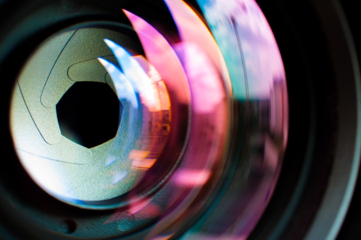
Much like the camera the iris is made to focus with contraction. Muscle iris is made up of pigment and fibers around the pupil. The pigment is called melanin. The more melanin there will be darker the color. The iris is protected behind the cornea. The entire eye is covered by a conjunctive layer. No two irises are the same. This is our identity.
Where do the light receptors take the light after it has been transferred into nerve impulses to the optic nerve (optic disk)? The part of the brain that is responsible for receiving light is the pituitary gland. Rests at the top of the spinal column under the brain’s center. The midbrain central location at the center of a function to our equilibrium testing along the sides is the sympathetic and parasympathetic nervous system. Occipital lobe is responsible for visuals. Light bounces off of random objects and catches our eye by focusing with the lens by ciliary body ligaments. The cells of the eyes capable of transmuting distance color are cone’s imagery. Light penetrates through retinas, layers of cells. Passing the Ganglion layer of cells and the Bipolar cells to reach the cones generate electrical impulses sure of colors. Generates aside from the optic disk from a focal point from the fovea centralis. The image should be beyond the retina, if it generates before the retina, the answer is convex or concave lens from the optical labs.
Our eyes are generally self cleansing. Lacrimal apparatus is the production of tears and drainage to the nasal cavity by lacrimal sac. Above the lateral canthus, where the outer corner of the eye’s eyelids meet is the lacrimal gland is under the bone of the eyebrow. With the help of excretory ducts these tears help the eye stay moist. As meibomian glands, under the eyelashes, produce an oil to prevent the eyelids from clinging together.
The fibrous layer of the eye is the sclera and cornea covered by a conjunctive layer. This is the outermost parts of the eye. Sclera is the white of the eye that is met with the optic nerve at the back of the eye, beyond its bony orbits. The very front of the eye that perceives refractions of light is the cornea. An open cavity of the eye that is clear between the cornea and the lens is called anterior chamber. Technically the five layers to the sclera, any indications of this change in hue could vary as emerges to passing, halt and of needed treatment.
Beneath the sclera is the choroid containing the blood vessels is the vascular layer. Another set of muscles to the organ that adjust the lens to near or far sighted. When the ciliary contracts the lens focuses at micro distances narrowing the lens. Three sets of suspensory ligaments pick up slack and change the focus. The fibers that make up the ciliary body lay longetidual, circular and radial collectively called zonular fibers.
Creating the pupils aperture of the iris. A muscle made of circular fiber and radial fibers each consisting of smaller fibers collectively making the Iris. Circular fibers are grouped from the sphincter pupillae and parasympathetic causing constriction. Dilation fibers consist of dilator pupillae and sympathetic. Also dilation usually depends on the amount of lighting inducing aperture. Pulses indicate a few central locations in the body.
The neural layer retina is full of photoreceptors (photons). Located below the pigment melanin is the non-visual retina. The optic part of the retina seen during ophthalmology shows the macula located next to the optic nerve (blind spot). A depression in the macula known as the fovea centralis, in this area of the eye contains the most photons that transfer light to electromagnetic pulses that travel to the optic nerve.
The extraocular muscles tells us about seven muscle attachments to the eye that give it names for it’s directional pulls. Also the cranial nerves that innervate the movements.
This is the anatomy and physiology of eye chemistry.
About the Creator
Enjoyed the story? Support the Creator.
Subscribe for free to receive all their stories in your feed. You could also pledge your support or give them a one-off tip, letting them know you appreciate their work.


Comments
There are no comments for this story
Be the first to respond and start the conversation.