
A novel imaging method enables researchers to examine the interior of living cells to better understand how cancer develops and spreads.
Observing the progression of cancer

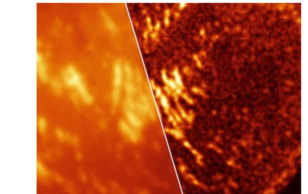
A novel class of fluorescent dyes has been discovered by researchers that allow for high-resolution cancer imaging in living cells, previously only possible with fixed samples. By revealing molecular behaviour that was previously concealed during live imaging, the method shows how cancer-related processes develop in real time.
Fluorescent dyes for imaging cancer
Tiny grip structures that develop and release during motion are visible at the border of a moving cell. Dr. Catherine Galbraith, an associate professor of biomedical engineering at OHSU, linked certain dye behaviours to the circumstances that allow these structures to be seen within living cells.
The findings demonstrated that local crowding, acidity, and cell motility can all affect how the same cellular characteristics look. Because of this variability, before applying the method to larger cellular systems, each dye must be properly matched to the biological context.
Why the new dyes are superior
For a straightforward reason, ordinary super-resolution microscopy—imaging that overcomes the typical blur of light—often fails in living cells. Cells may be stressed by systems that rely on strong light or other chemicals to turn dyes on and off.
This stress is important, particularly in cancer biology, where researchers require minutes of behaviour rather than a single frozen picture. The new dyes eliminated most of that setup overhead by blinking naturally, making advanced imaging accessible to more common labs.
Not a single victor
Researchers now have a panel rather than a single favourite thanks to chemical adjustments that pushed on-off ratios across two orders of magnitude. While some versions handled sparser targets or faster image sequences, others performed better when molecules were closely packed together.
Lower on-off ratios typically resulted in clearer molecular pinpoints in fixed cells, but glow timing in live cells upset the equilibrium. For this reason, the study advocates for a set selected by context rather than a one-size-fits-all label.
Observing the progression of cancer
Focal adhesions, protein-rich grasp sites that aid cells in crawling through tissue, may be the most stunning observation for cancer researchers. As cells push forward, those structures develop and disintegrate; this process is linked to invasion and the spread of cancer.
The National Cancer Institute (NCI) views cell migration as a fundamental component of cancer spread because metastatic tumour cells must migrate and penetrate surrounding tissue. According to Galbraith, "these tools allow us to see critical cancer-related processes happening live, at the very small scale where key decisions are made inside cells."
After the activation of genes
The dyes also monitored chromatin, which is DNA encased in proteins, inside the nucleus while cells were still living and undergoing changes. That was significant since the degree to which this material is packed closely and whether crowded areas open affects gene activity.
Additionally, one variant tracked how open DNA remained while producing six times as many useful molecular detections as an earlier light-activated label. These improvements make it simpler to monitor gene-control activity without freezing cells all at once.
Acidity affects dyes.
It turns out that acidity was so important that a dye that worked well in one compartment might not work well in another. One low-glow dye brightened images in lysosomes, which are acidic recycling compartments inside cells, because acid altered the dye's blinking behaviour.
By neutralising the compartment, fixing the cells undid that benefit, and a new dye then worked better. This environmental sensitivity may be helpful rather than problematic in cancer research because tumours frequently contain acidic pockets.
Paying attention to RNA messages
When the team combined the blinking dyes with RNA labelling probes, messenger RNA—genetic instructions that transmit DNA's code to produce proteins—also showed up as more distinct signals. This made it easier for the microscope to distinguish between adjacent RNA signals, enabling researchers to determine whether genes function independently or in groups.
Quick readouts were made possible by a second imaging mode that used less than 1,000 frames to create clearer images from brightness flickers. That speed can expand the scope of standard research for labs monitoring RNA while cancer cells switch genes on and off.
The importance of resolution and speed
When the researchers monitored expanding cell attachments and moving mitochondria over time, speed was just as important as sharpness. One method exchanged some detail for quicker updates, while another tracked individual light molecules for the greatest detail.
One live-cell arrangement operated three times faster than previous work, and the flicker-based method visualised minuscule new attachments near the front border. The findings imply that if researchers can match resolution and time to biology, they don't require a single ideal method.
Using the dyes for imaging cancer
Since many cancer labs lack specialised equipment or the patience to fine-tune images, the ease of usage may determine whether these dyes are important. According to Galbraith, "new tools open up biology." Nevertheless, because the incorrect dye may perform poorly when density, acidity, or fixation vary, the panel is not plug-and-play.
The OHSU team has effectively given researchers useful guidelines, which could accelerate acceptance. The new dyes increased the versatility of live-cell microscopy, from moving cell borders to packed DNA and acidic vesicles.
About the Creator
Enjoyed the story? Support the Creator.
Subscribe for free to receive all their stories in your feed.
Comments
There are no comments for this story
Be the first to respond and start the conversation.