
The Cardiovascular System- The Heart.
Heart


Introduction
The human body is made of many systems that work together to ensure the normal functioning of an individual; the cardiovascular system is responsible for transporting oxygenated blood from the lungs to the heart and from the heart together with other important substances to the rest of the body. Deoxygenated blood and other substances such as carbon dioxide are transported from all parts of the body to the heart and then the lungs; the circle then repeats.
The Cardiovascular System:
It is the system that is mainly responsible for transporting oxygenated and blood rich in nutrients from the lungs to the left auricle of the heart and then to the left ventricle and cells of the body and transporting deoxygenated blood from cells of the body to the right auricle, to the right ventricle, and then to the lungs. The main components of the cardiovascular system are the heart, blood, and blood vessels.
The Blood Vessels
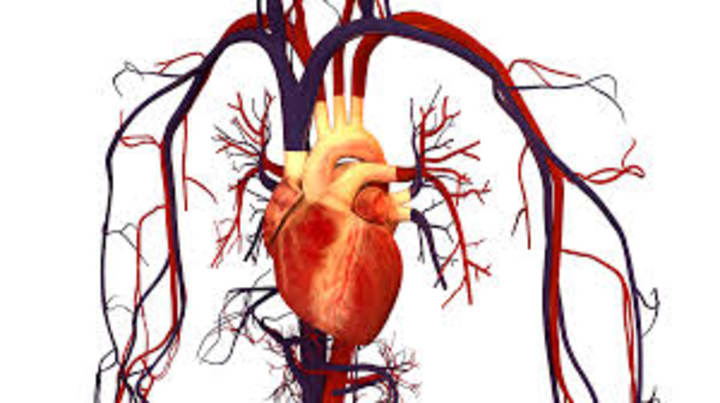
These are structures through which blood travels in the body; there are four main types of blood vessels: the aorta, arteries, veins, and capillaries. The superior vena cava drains blood from the upper part of the body above the heart into the right auricle while the inferior vena cava drains blood from the lower part of the body below the heart into the right auricle.
The Aorta: it is the largest blood vessel found in the human body; it transports oxygenated blood from the heart to the arteries under high pressure.
The Artery: it is the second largest blood vessel found in the body; it branches from the aorta and is found deeper within the skin- because it transports blood under high pressure from the left ventricle- compared to that of veins and capillaries. They transport oxygenated and blood rich in nutrients; they branch into capillaries. The pulmonary artery is the only artery that transports deoxygenated and blood-poor nutrients from the heart to the lungs.
The Capillary: it is the smallest blood vessel with tin walls listed among the four blood vessels; the main function of capillaries is the exchange of gases, nutrients, etc., in tissues, cells, and organs.
The Vein: it is the third largest blood vessel found in our body; the function of veins is to transport deoxygenated blood and other waste substances from cells to the right auricle, right ventricle, and the lungs. The pulmonary vein is the only vein that transports oxygenated blood from the lungs to the left auricle for circulation.
THE HEART
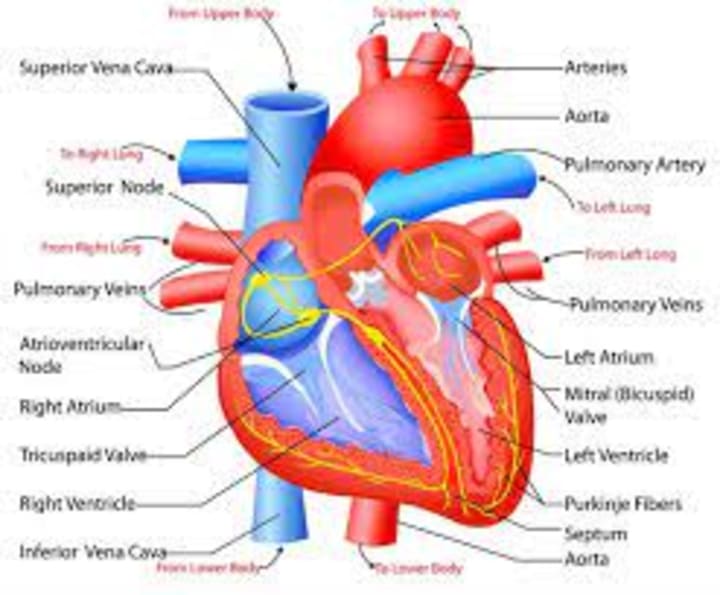
The heart is a pear-shaped organ made of muscles called cardiac muscles that are located in the chest cavity a little to the left. It is made of four chambers- the right and left auricles and the right and left ventricles-, valves- the aortic valves, tricuspid valves, bicuspid also called the mitral valves, and the pulmonary valves-; extending from the top of the heart is the aorta which branches into the aortic arch. Three blood vessels extend from the top of the aortic arch; the coronary artery supplies the heart muscles with oxygenated blood rich in nutrients. The septum separates the heart into two, the right hemisphere and the left hemisphere.
The heart is a unique structure that generates its electrical impulses which maintain a normal heartbeat; two main structures are responsible for generating and maintaining heartbeats: the sinoatrial (SA) nodes and the atrioventricular (AV) nodes. Electrical impulses are generated within the wall of the right auricle and are propagated through the His- Purkinje conduction system that stimulates the contraction of the ventricles.
HEART BEAT
It is the number of times a heart beats within a minute; it is normally from 60 to 100 beats per minute for adults and more for children. Regular heartbeats can be disrupted- a condition called arrhythmia- due to certain conditions that cause the SA node to irregularly transmit electrical impulses.
A defibrillator is an electrical device that resets an irregular heartbeat by stopping the heart completely and allowing the SA node to take over the regular transmission of electrical impulses enabling contraction and relaxation of the heart muscles.
THE FLOW OF BLOOD THROUGH THE CARDIOVASCULAR SYSTEM
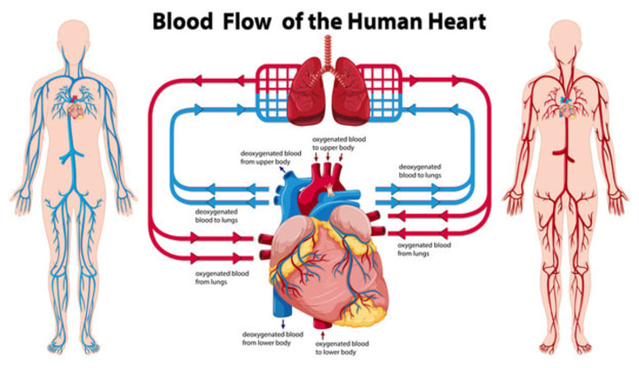
There are two main types of blood circulation: pulmonary circulation and systemic circulation.
Pulmonary Circulation: it is the flow of oxygen-poor blood from tissues, cells, and organs to the lungs where gaseous exchange- the removal of carbon dioxide from the blood and the uptake of oxygen by the blood- takes place. The pulmonary artery is the only artery that transports deoxygenated blood in the body.
Systemic Circulation: it is the flow of blood rich in oxygen and other important substances from the left ventricle to tissues, cells, and organs of the body.
IMPORTANT STRUCTURES OF THE HEART AND THEIR FUNCTIONS
The major structures of the heart are the epicardium, myocardium, endocardium, pericardium, septum, right and left ventricles, right and left auricles, the tricuspid valves, mitral or bicuspid valves, aortic valves, and pulmonary valves.
The Epicardium: it is the outermost covering of the heart mainly made of cardiac muscles; coronary arteries supply cardiac muscles with blood rich in oxygen.
The Myocardium: it is the muscular middle layer of the heart.
The Endocardium: it is the innermost portion of the heart.
The Pericardium: it is a fluid-filled sack that surrounds the outer covering of the heart.
Septum: it is a muscular layer that separates the heart into two halves- the right and left chambers.
The Right Auricle: deoxygenated blood from the upper and lower parts of the body empties into the right auricle. The superior vena cava empties blood from the upper part of the body above the heart while the inferior vena cava empties blood from the lower part of the body into the right auricle; blood from the right auricle empties into the right ventricle.
The Right Ventricle: the right ventricle is less muscular because it pumps deoxygenated blood through a short distance to the lungs for oxygenation.
The Left Auricle: oxygenated blood from the lungs empties into the left auricle and then the left ventricle.
The Left Ventricle: it is the most muscular chamber of the heart because it pumps oxygenated blood farther away from the heart to cells, tissues, and organs.
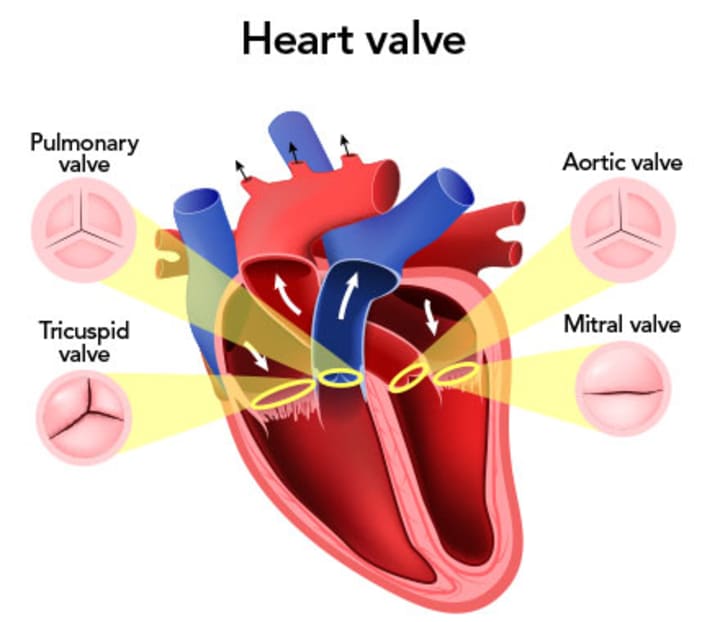
The Tricuspid Valves: These are three sets of valves located between the right auricle and right ventricle; they direct the one-way flow of blood into the right ventricle. The purpose of the heart’s valves is to prevent the backflow of blood allowing blood to flow in only one direction.
The Pulmonary Valve: it is located in the pulmonary artery above the right ventricle and prevents the backflow of deoxygenated blood into the right ventricle.
The Bicuspid/Mitral Valves: they are two sets of valves located between the left auricle and left ventricles; they allow the one-directional flow of oxygenated blood from the left auricle into the left ventricle.
The Aortic Valves: These are valves that are located in the aorta above the left ventricle; they prevent the backflow of oxygenated blood from entering the left ventricle.
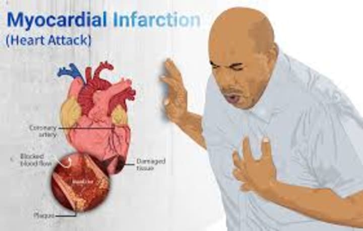
Heart Attack
The main cause of heart attack is when heart muscles do not receive the required oxygen and nutrients needed for them to function properly; when this occurs over a period, heart muscles starve and stop working resulting in a heart attack. The coronary artery innervates the heart supplying it with blood rich in oxygen; when there is obstruction in the flow of blood to a particular region of the heart, cardiac muscles in that region begin to die leading to improper heartbeats which then result in a heart attack. These obstructions are caused by a blockage in the coronary artery by calories, fat, blood clots, and low-density lipoprotein (LDL) which is bad cholesterol.
This is also the main cause of stroke- when a region of the brain does not receive an adequate supply of oxygenated blood rich in nutrients, cells in that region of the brain die causing that region of the brain to not function properly leading to stroke.
Conclusion: The heart is a complex and delegated organ; the information above is the basics to knowing and understanding the basic functions of the heart, and knowing important parts of the heart, etc. It should be noted that some diseases and complications are associated with the heart, and we should put all safety measures in place to maintain a healthy heart because each of us has just one heart. The heart is delicate such that nature positioned it in a protective cage called the rib cage with the sternum to support and strengthen its structure.
Author: Sekou Sesay


About the Creator
Sekou Sesay
Love
Keep reading
More stories from Sekou Sesay and writers in Education and other communities.
THE STRUCTURE AND FUNCTION OF PARTS OF A FLOWER.
Introduction: The flower is the most attractive part of most plants; flowers that are pollinated by animals have brightly colored petals and those pollinated by wind have less brightly colored petals. Flowers undergo two types of pollination referred to as cross-pollination and self-pollination; most flowers that undergo self-pollination are referred to as perfect flowers because they have both male and female sex organs; and some flowers that undergo cross-pollination have one sex organ responsible for reproduction and they are referred to as imperfect flowers.
By Sekou Sesay5 months ago in Education
Top 10 Benefits of Outsourced CFO Service to Increase Profits
What are outsourced CFO services? Outsourced CFO (Chief Financial Officer) services refer to the practice of hiring a professional financial expert or a team of experts who work remotely to provide financial services and advice to businesses. These services can include managing financial risks, creating budgets, forecasting financial trends, analyzing financial data, providing financial reports, and creating strategic plans to improve financial performance.
By Chirag koshti3 days ago in Education


Comments
There are no comments for this story
Be the first to respond and start the conversation.