
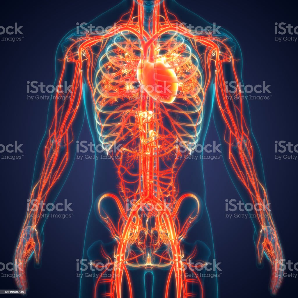
The circulatory system, a crucial part of the overall circulatory framework, is responsible for the circulation of blood. It encompasses the cardiovascular system and the lymphatic system, where lymph is transported. The terms "circulatory system" and "cardiovascular system" are often used interchangeably to describe the process of blood circulation. The cardiovascular system comprises the heart, blood, and blood vessels. Acting as a pump, the heart propels blood through these vessels. With two sides, each housing two chambers, the heart is structured.
The primary role of the circulatory system is the transfer of inhaled oxygen from the lungs to the body's tissues, coupled with the removal of carbon dioxide, which is then exhaled. Essentially, oxygen-depleted blood from the body returns to the right side of the heart, from where it is directed to the lungs. Within the lungs, blood acquires oxygen and releases carbon dioxide. Subsequently, oxygen-enriched blood returns to the left side of the heart, a component known as the pulmonary circuit. This oxygen-rich blood is then pumped to the body's tissues by the left side of the heart, where it releases oxygen and accumulates carbon dioxide. This phase is termed the systemic circuit. Given its responsibility to distribute blood to the entire body, the left side of the heart boasts thicker muscular walls than the right side.
Four valves ensure unidirectional blood flow through the heart: oxygen-poor blood flows from the right atrium to the right ventricle and then to the pulmonary arteries, while oxygen-rich blood moves from the left atrium to the left ventricle and subsequently into the aorta. Shielding the heart is a dual-layered protective sac called the pericardium. This sac's pericardial cavity houses fluid that serves as a lubricant, facilitating the heart's contraction and relaxation with minimal friction. The heart wall consists of three layers: the outer epicardium, lining the pericardial cavity; the inner endocardium, lining the heart chambers and valves, which connects to the endothelium of blood vessels; and the robust myocardium, the muscular tissue driving the heart's rhythmic beating.
Initiated by electrical impulses referred to as action potentials, the contraction of the heart muscle is self-generated, unlike skeletal muscles that require stimulation from the nervous system. These impulses emanate from pacemaker cells, a group of cells constituting the cardiac conduction system. The principal pacemaker is the SA node, responsible for initiating all heartbeats and regulating heart rate. Beyond its role in gas transport, blood is pivotal in supplying nutrients to body tissues and eliminating metabolic byproducts. Nutrients are obtained from the digestive system, absorbed through the small intestine's walls into the bloodstream. The liver processes these substances for toxins before they join the general circulation.
Within tissues, nutrients are exchanged for waste products, which are subsequently filtered by the kidneys and expelled as urine. Furthermore, blood transports hormones from endocrine glands to target organs, playing a pivotal role in the body's immune defense. Blood is comprised of two primary components: plasma, a clear extracellular fluid, and formed elements such as red blood cells, white blood cells, and platelets. Red blood cells are responsible for oxygen and carbon dioxide transport, white blood cells participate in defense mechanisms against pathogens, and platelets are integral to blood clotting, reducing blood loss during injuries.
Operating as a closed loop, the blood circulatory system ensures blood remains within the vessels at all times. Substances move to and from the surrounding tissues through the walls of blood vessels via diffusion. Vessels that carry blood away from the heart are termed arteries, while those returning blood to the heart are referred to as veins. Typically, arteries transport oxygenated blood, whereas veins carry deoxygenated blood. However, for pulmonary arteries and veins, this arrangement is reversed. The conventional path of blood flow involves the heart propelling blood into large arteries, subsequently smaller arteries, arterioles, the tiniest blood vessels known as capillaries, facilitating substance exchange. Blood then converges into small veins (venules), proceeding through larger veins and returning to the heart.
Arteries and veins serve the function of conveying blood, consisting of three layers: the outer layer composed of loose connective tissue anchors the vessels, the middle layer of predominantly smooth muscle permits constriction or dilation to regulate blood flow, and the inner layer consists of thin squamous endothelium separated from outer layers by a basement membrane. Generally, larger vessels exhibit more connective tissue and smooth muscle. Arteries exhibit more muscular tissue than veins due to their role in transporting blood away from the heart, which generates higher pressures. Capillaries, responsible for exchanging substances between blood and surrounding tissue, solely consist of a thin endothelium and its basement membrane, facilitating the diffusion of blood solutes.
About the Creator
Enjoyed the story? Support the Creator.
Subscribe for free to receive all their stories in your feed. You could also pledge your support or give them a one-off tip, letting them know you appreciate their work.
Keep reading
More stories from ackun vlog and writers in Education and other communities.
The Respiratory System
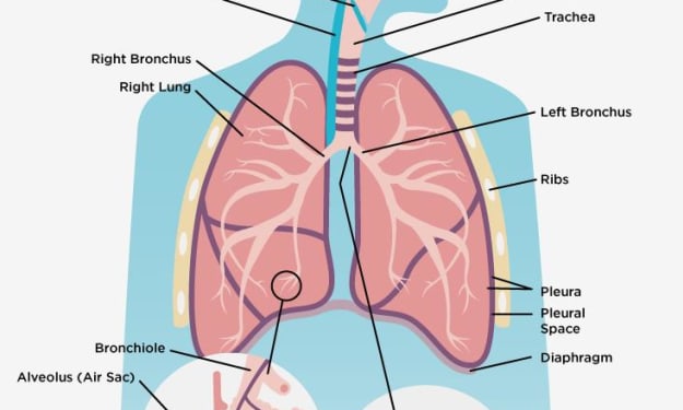
THE RESPIRATORY SYSTEM The respiratory system consists of all the organs involved in breathing, including the lungs and airways. The two most important functions of the respiratory system are to provide us with oxygen and to rid our bodies of carbon dioxide. The respiratory system has two tracks: upper and lower. The upper track is above the sternal angle, outside of our thorax, and above the level of our vocal cords. This upper track helps to moisten air before it enters our lungs. Within this track we have three parts: the nasal oral cavity, the pharynx, and the larynx. The nasopharynx is where air enters our mouth or nose, the oropharynx is where air enters our mouth from below, and the lorenzo pharynx connects to our esophagus. The laryngeal pharynx plays an important role in allowing us to speak and is connected to the larynx.
By ackun vlog10 months ago in Education
TV advertising revenue
The Early Days In the early days of television, advertising was a fledgling industry, much like TV itself. The first TV commercial aired in 1941, when the Bulova Watch Company paid $9 for a 10-second spot during a baseball game broadcast on WNBT in New York. This simple black-and-white ad marked the beginning of a new era in advertising, where moving images and sound could be used to promote products and services directly to consumers in their homes.
By m habibullah5 days ago in Education
"Empowering Women in Education"
In the realm of education, the quest for gender equality has been both a journey and a destination. Across the globe, efforts to empower women in education have aimed not only to close the gender gap but also to foster a more inclusive and equitable society. From grassroots initiatives to policy reforms, various approaches have been employed to ensure that women and girls have equal access to educational opportunities and are encouraged to pursue their academic and professional aspirations without barriers.
By Dharanidharan T 5 days ago in Education


Comments
There are no comments for this story
Be the first to respond and start the conversation.