
THE RESPIRATORY SYSTEM
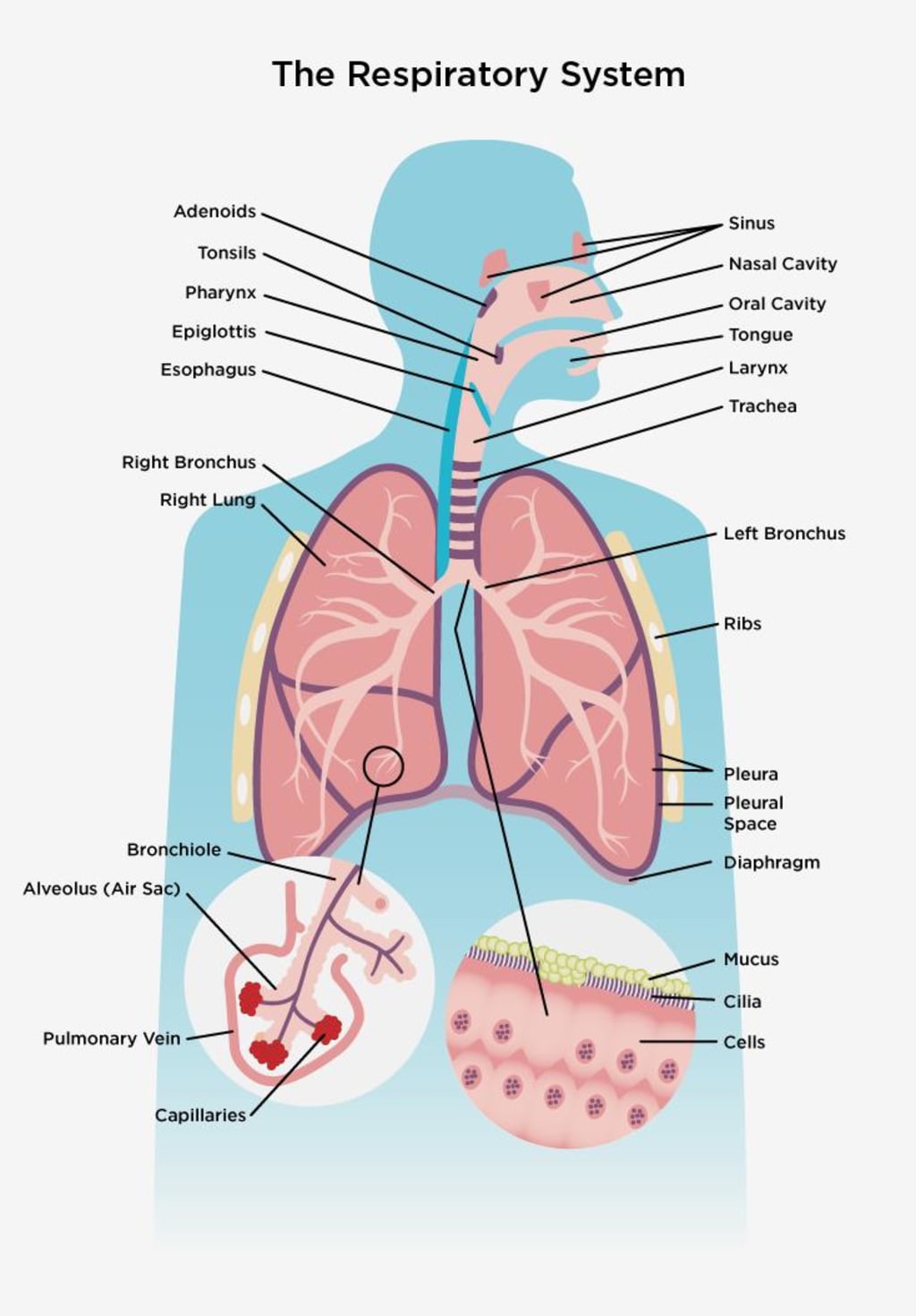
The respiratory system consists of all the organs involved in breathing, including the lungs and airways. The two most important functions of the respiratory system are to provide us with oxygen and to rid our bodies of carbon dioxide. The respiratory system has two tracks: upper and lower. The upper track is above the sternal angle, outside of our thorax, and above the level of our vocal cords. This upper track helps to moisten air before it enters our lungs. Within this track we have three parts: the nasal oral cavity, the pharynx, and the larynx. The nasopharynx is where air enters our mouth or nose, the oropharynx is where air enters our mouth from below, and the lorenzo pharynx connects to our esophagus. The laryngeal pharynx plays an important role in allowing us to speak and is connected to the larynx.
Here in this region, we find a fold of connective tissue, visible as the epiglottis, which closes to shield the airway, preventing aspiration. Moving on, we delve into the final segment of the upper respiratory tract known as the larynx, commonly referred to as the voice box. While it's occasionally categorized as part of both the upper and lower respiratory tracts, it plays a crucial role in both air passage and vocalization. The vocal folds, prominently displayed here, stand as key landmarks for intubation.
Transitioning to the lower respiratory tract, we confront a more intricate landscape. Often denoted as the respiratory or tracheobronchial tree due to its branching nature, this intricate structure unveils its complexity. Within this lower realm, four main segments stand out. The initial component is the trachea, a cartilaginous tube connecting the larynx and lungs. It boasts complete cartilage rings at its upper end, the cricoid cartilage, while its lower end is characterized by incomplete C-shaped rings of elastic hyaline cartilage. The tracheal lining features ciliated epithelial cells housing goblet cells that synthesize mucins, pivotal in mucus production. Narrowing or blockage here results in stridor, the distinct high-pitched sound accompanying breathing, primarily during inspiration.
Shifting our focus to the second portion, we encounter the bronchi, marking the commencement of lower respiratory tract segmentation. Within this realm, we classify the primary bronchi into left and right main bronchi, distinguished by the Carina, the point of tracheal division near the fifth vertebra. These bronchi serve as conduits for air to each lung, with the right bronchus being wider, shorter, and more vertically oriented, potentially posing a direct route for aspiration or endotracheal tube insertion complications. Both the left and right bronchi possess expansive lumens, lined by ciliated epithelial cells and accompanied by underlying smooth muscle cells equipped with mucous-producing capabilities.
As we trace the course of branching within this system, it becomes evident that hyaline cartilage gradually diminishes while smooth muscle content increases in the airways. This interplay of muscle and cartilage serves the crucial purpose of maintaining airway patency. Beyond the primary bronchi, the journey continues with secondary bronchi, also known as lobar bronchi. These segments further divide to direct air to specific lung lobes. Accordingly, three secondary bronchi branch from the right primary bronchus, while the left primary bronchus gives rise to two, aligning with the left lung's two lobes.
Progressing onward, we encounter tertiary bronchi, often termed segmental bronchi, responsible for channeling air to each segment within a lung lobe. The branching system persists with sub-segmental bronchi, representing the fourth, fifth, and sixth orders of division. This intricate pattern leads us to bronchioles, the smaller, more delicate branches of the respiratory tract. Unlike their larger counterparts, bronchioles lack hyaline cartilage support and instead rely on elastic fibers linked to surrounding lung tissue. These bronchioles undergo multiple levels of branching, culminating in terminal bronchioles. These terminal bronchioles mark the conclusion of the conducting bronchioles and herald the transition to the respiratory zone.
Within this respiratory zone, we find the respiratory bronchioles, the gateway to the final frontier of the lower respiratory tract – the lungs. Here, the alveoli come into play, these microscopic air sacs facilitating the all-important gas exchange process. This intricate system commences with respiratory bronchioles, followed by alveolar ducts that bridge the gap between these bronchioles and the alveolar sacs. The alveolar sacs, in turn, house individual alveoli, the sites where oxygen and carbon dioxide exchange transpires. Remarkably, the human lung boasts a staggering 300 to 500 million alveoli, collectively furnishing a massive surface area exceeding 750 square feet (or approximately 70 square meters).
Each alveolus is enveloped by a network of capillaries that cover around 70% of their surface area, promoting efficient gas exchange. While the mechanics of gas exchange are more intricate, I've created another resource dedicated to this topic, which you can access for more detailed insights. The culmination of this intricate system lies within the lungs, the hub where the vital exchange of gases transpires.
Shifting our focus, let's delve into the muscles that orchestrate respiration. These muscles, grouped into three primary categories, are integral to the mechanics of inhalation and exhalation. Leading the ensemble is the diaphragm, a domed muscle lying at the thoracic-abdominal interface. Its contraction during inhalation expands the thoracic cavity, drawing air into the lungs. Upon exhalation, the diaphragm relaxes, allowing elastic recoil to compress the thoracic cavity, expelling air. Complementing this is the intercostal muscle group, nestled between the ribs, orchestrating ribcage movement to further facilitate inhalation. The third group, accessory muscles, comes into play during heightened demands like exercise, supplementing respiration efforts.
Finally, let's explore the pulmonary vasculature. This system comprises two arterial circulations within the lungs – the pulmonary circulation and the bronchial circulation. In the former, pulmonary arteries carry deoxygenated blood to the lungs for gas exchange, while pulmonary veins return oxygenated blood to the left atrium. This low-pressure system is designed to optimize gas exchange. On the other hand, the bronchial circulation, fed by systemic arteries, supplies oxygenated blood to lung tissue. This high-pressure system is essential for lung nourishment. An intriguing facet is the creation of a physiological shunt, as a portion of deoxygenated blood from the bronchial circulation mixes with oxygenated blood, returning to the left heart. This multifaceted vascular network completes the remarkable respiratory system.
Remember that this overview provides a comprehensive understanding of the respiratory system's intricacies. For a more detailed exploration of specific aspects, such as gas exchange, I recommend referring to supplementary resources. If you found this overview helpful, please consider liking and sharing it.
About the Creator
Enjoyed the story? Support the Creator.
Subscribe for free to receive all their stories in your feed. You could also pledge your support or give them a one-off tip, letting them know you appreciate their work.
Keep reading
More stories from ackun vlog and writers in Education and other communities.
The Nervous System
This morning unfolded in the usual manner for me. I awoke with thoughts lingering about the recurring dream involving the guy in the sloth costume. Afterward, I got dressed as the chill in the air demanded it. Following that, I prepared some toast with butter due to my hunger, and I attended to my dog's needs, as she was whining and gazing at me. Next, I brewed some tea, allowing it to cool off before consumption to prevent a repeat of the mouth burn from yesterday.
By ackun vlog10 months ago in Education
Top Technologies Transforming Construction Site Security in 2024
In the dynamic landscape of construction, security remains a paramount concern. With the advancement of technology, the industry witnesses a significant transformation in how construction sites are safeguarded. In 2024, innovative solutions are revolutionizing construction security, providing unprecedented levels of protection against various threats. This article explores the top technologies shaping the future of construction security and their implications for the industry.
By Alpine protection services4 days ago in Education
AI Sentence Generator
An AI sentence generator is a sophisticated tool designed to produce human-like text based on given input prompts. This technology leverages advanced algorithms and machine learning techniques, particularly natural language processing (NLP), to understand context, semantics, and linguistic patterns. The development of AI sentence generators involves training models on extensive datasets comprising diverse text sources, enabling them to generate coherent and contextually relevant sentences.
By AI Sentence Generator3 days ago in Education


Comments
There are no comments for this story
Be the first to respond and start the conversation.