To peer Inside the Nanoworld: Let’s Put Electron Microscopy to Address Major Challenges in Material Science Industry
Electron microscopes are one of the most powerful tools capable of distinguishing even individual atoms. How do electron microscopes allow detailed investigations of materials? What can we do with electron microscopy studies?

1. What is Electron Microscopy?
Electron microscopy (EM) has practical applications in a wide range of fields, including materials science, microelectronics, biology, environmental science, and geology. Techniques based on EM are used to probe the structure and composition of materials, providing an understanding of their properties and behaviour.
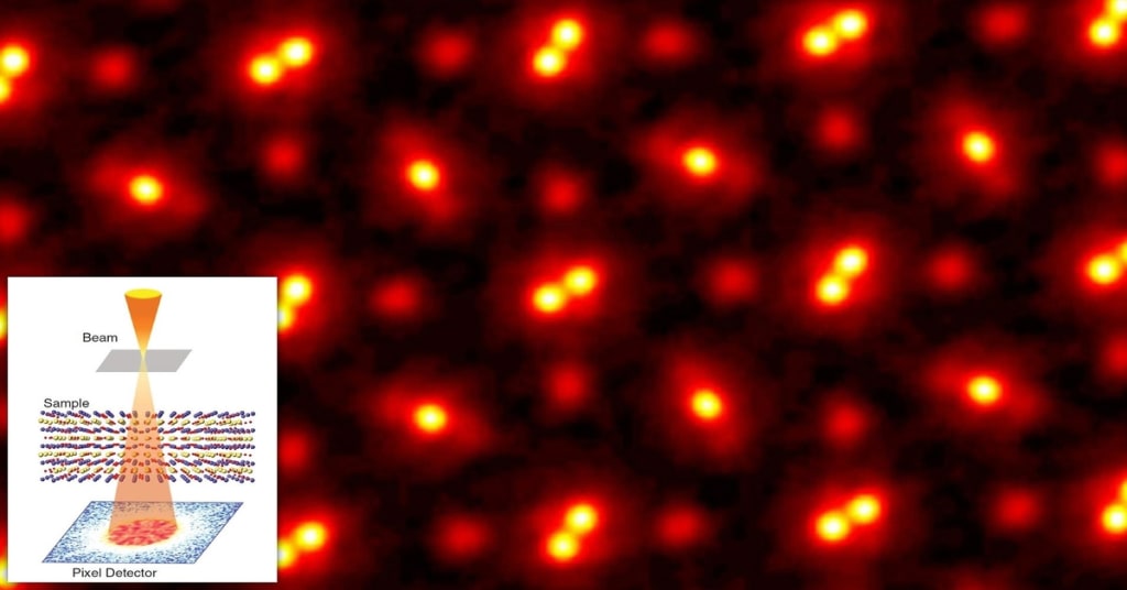
Unlike light microscopes, which use visible light to produce images, electron microscopes use a beam of electrons. When it comes to magnifying nanoscale materials, electrons are better than visible light. That’s because electrons, which have wave-like properties due to quantum mechanics, have wavelengths a thousand times shorter than photons. Shorter wavelengths produce a higher resolution, much like a finer thread can create more intricate embroidery. This allows electron microscopes to achieve much higher resolution and magnification than light microscopes, magnifying micrometre and nanometer objects by up to ten million times. These high-resolution EM analyses provide a spectacular level of detail, allowing researchers to view single atoms. In a typical electron microscopy setup, a sample is placed in a vacuum chamber, and a beam of electrons is generated and focused onto the sample using a series of electromagnetic lenses. The electrons interact with the sample, and the resulting signals are detected to produce an image of the sample.
2. EM Techniques and Potential Information about a Material
2.1 High-Resolution Imaging: Electron microscopy imaging techniques use a beam of electrons to create high-resolution images of samples at the nanoscale level. It is particularly useful for studying the structure and properties of materials and biological samples at a very small scale. The EM images can provide detailed information about a sample’s shape, size, and arrangement of atoms. Transmission electron microscopy (TEM) images are created by passing a beam of electrons through a thin sample and collecting the transmitted electrons on a detector on the other side. The contrast in the image is generated by the difference in the number of electrons that are transmitted through different regions of the sample. The first TEM method capable of the atomic resolution was high-resolution (HR) TEM imaging. In HRTEM, a coherent parallel electron beam elastically interacts with the materials’ crystalline lattice, resulting in an interference pattern (HRTEM image) that has the same periodicity as the crystal and can be used to retrieve information on symmetry. The development of aberration-corrected TEMs has enabled imaging of the materials with a higher resolution, even in picometers (pm).
2.2. Understanding the Crystallinity of Materials: EM provides unique opportunities for crystal structure analysis at a very local scale complementing powder diffraction techniques, such as X-ray or neutron powder diffraction. A deep understanding of the relationship between the properties and the crystal structure is the key to designing new materials and improving existing ones. Selected area electron diffraction (SAED) is a TEM technique to obtain diffraction patterns that result from the electron beam scattered by the crystalline sample. In SAED, the elastically scattered electron from the crystal lattice obeys Bragg’s law; therefore, the diffraction pattern is completely dependent on the interplanar spacings (dhkl) and composition of the crystal being analyzed. Electron diffraction (ED) patterns contain information on the symmetry of the crystal, and, in some cases, the intensities of the reflections can be used quantitatively to solve the crystal structure.
2.3. Probing Composition of Materials: Coupling of scanning transmission electron microscopy (STEM) with atomic resolution spectroscopic techniques, such as energy-dispersive X-ray spectroscopy (EDS) and electron energy loss spectroscopy (EELS), allows analysis of the chemical composition oxidation state, and coordination number of the individual atomic columns in the structure. When the materials are sufficiently stable under the electron beam, the diffraction patterns and images can be complemented with high-resolution EDS or EELS maps, allowing to support the structure models with direct knowledge of the distribution of the elements over the different solved atomic positions. High-angle annular dark field (HAADF)-STEM imaging is a standard technique for the structural and compositional characterization of different nanomaterials at the atomic scale. By studying the composition of materials using electron microscopy, researchers can better understand the material’s properties and behaviour. For example, they can identify the presence of impurities and defects and understand their impact on the material’s performance.
3. Implications of Electron Microscopy Analyses in a Few Industries
3.1. Materials Syntheses: Electron microscopy is a powerful tool that is used extensively in materials science to study the structure and composition of materials at a very small scale. By studying the microstructure of materials using EM, researchers can probe morphology (size, shape), defects, compositions, purity, elemental distribution, and crystallinity of materials. This information can be used to optimize the manufacturing process and improve the properties of the materials (e.g., strength, conductivity, and durability).
3.2. Microelectronics: In the semiconductor industry, EM is used to inspect the surface of microchips and other microelectronic devices to ensure their quality and performance. The high magnification and resolution of electron microscopes allow for the detailed analysis of the materials and structures used in microelectronic devices, enabling researchers to make improvements to the manufacturing process.
3.3. Batteries and Renewable Energy Materials: Electron microscopy can be used in energy research to study the microstructure of materials used in renewable energy technologies, such as solar cells, catalysis, and batteries. By using high-resolution TEM, researchers can observe the detailed structure of these materials, including their grain size and defects, and understand their impact on the performance of the material. This information can be used to optimize the manufacturing process and improve the performance of renewable energy technologies. In-situ TEM analyses can also be used to study the electrochemical processes that take place within batteries during charging and discharging, providing valuable insights into improving the battery’s efficiency and lifespan. This information can provide valuable insights into the performance of the battery and help to improve its design and manufacturing process. High-resolution electron microscopy can be used to identify defects in the materials used in the battery, allowing researchers to understand their impact on the battery’s performance and develop strategies to mitigate them.
3.4. Mining Industry: There are several ways that electron microscopy can be used in the mining industry. For example, electron microscopy can be used to study the microstructure of minerals and ores to determine their composition and quality. This analysis can help to identify valuable minerals and optimize the mining process. Electron microscopy can also be used to study the surface of mining equipment, such as drills and excavators, to identify wear and tear and develop strategies for improving their durability and performance. Additionally, electron microscopy can be used to study the environmental impact of mining operations, such as the presence of pollutants in soil and water samples.
3.5. Drug Discovery and Biology: Electron microscopy can be used in drug discovery to study the structure of viruses and bacteria at a very small scale. For example, cryo-TEM can provide high-resolution images of the internal structure of these microorganisms, allowing researchers to identify key proteins and other molecular targets for new drugs. By studying the structure of viruses and bacteria, researchers can develop drugs that specifically target these structures, reducing their ability to cause disease. Electron microscopy can also be used to study the structure of existing drugs and their interactions with viruses and bacteria, providing valuable insights into how to improve their effectiveness.
About the Creator
Rana Faryad
I hold a Ph.D. in Nanotechnology and write about exciting technologies to reshape the future of humanity.
Enjoyed the story? Support the Creator.
Subscribe for free to receive all their stories in your feed. You could also pledge your support or give them a one-off tip, letting them know you appreciate their work.


Comments
There are no comments for this story
Be the first to respond and start the conversation.