
The Journey of Visual Processing: Exploring the Marvels of the Visual Cortex
Science
Introduction:
In the fascinating realm of neuroscience, understanding how our brain processes information from our surroundings is a captivating endeavor. In this article, we will delve into the intricate process of visual processing, starting from the moment light interacts with our eyes to the interpretation of visual information by the brain. Join us on this journey as we explore the wonders of the visual cortex and the remarkable mechanism of visual transduction.
The Basics of Vision:
Vision relies on the interaction of light with our eyes. Visible light, a narrow band of electromagnetic radiation, ranging from 400 to 700 nanometers, enables us to perceive our surroundings. The human eye's intricate structure allows light to enter through the pupil, regulated by the iris, and be focused by the lens onto the retina. Binocular disparity, facilitated by having two eyes, provides us with depth perception.
The Retina: Where Light Meets Neurons:
The retina, located at the back of the eyeball, plays a vital role in converting light into neural signals. It consists of five layers of different types of neurons, including receptor cells (rods and cones), horizontal cells, bipolar cells, amacrine cells, and retinal ganglion cells. Rods and cones, found at the back of the retina, mediate scotopic and photopic vision, respectively, enabling us to perceive our surroundings under different lighting conditions.
Rods and Cones: Different Visions, Different Roles:
Rods and cones differ in their structure and function, mediating different types of vision. Cones are responsible for photopic vision in bright light, providing detailed perception, while rods enable scotopic vision in dim lighting conditions, sacrificing detail for enhanced sensitivity. The convergence of rods and cones onto retinal ganglion cells determines the level of sensitivity and acuity, allowing us to adapt to various lighting conditions.
The Fovea: The Center of Visual Acuity:
Within the retina, the fovea, a small indentation at its center, contains only cones. This specialization grants the fovea the ability to perceive small details with high acuity. Moving away from the fovea, the density of cones decreases, and rods become more prevalent, reaching their maximum density at approximately twenty degrees from the fovea.
Color Perception and Photoreceptors:
Color perception relies on three types of cones: red, green, and blue (also known as L, M, and S cones). Trichromatic color theory explains how the differential activation of these cones enables us to perceive a wide range of colors. Additionally, opponent process theory describes how the connections between cones and ganglion cells produce complementary color perceptions and negative afterimages.
Visual Transduction: From Light to Neural Signals:
Visual transduction occurs in the rods and cones through a protein called rhodopsin. Rhodopsin absorbs light and triggers a cascade of events that lead to the closure of sodium channels in the rods, hyperpolarizing them. This process reduces the release of inhibitory neurotransmitters, allowing bipolar cells to depolarize and transmit signals to retinal ganglion cells. These signals travel along the optic nerve to the primary visual cortex.
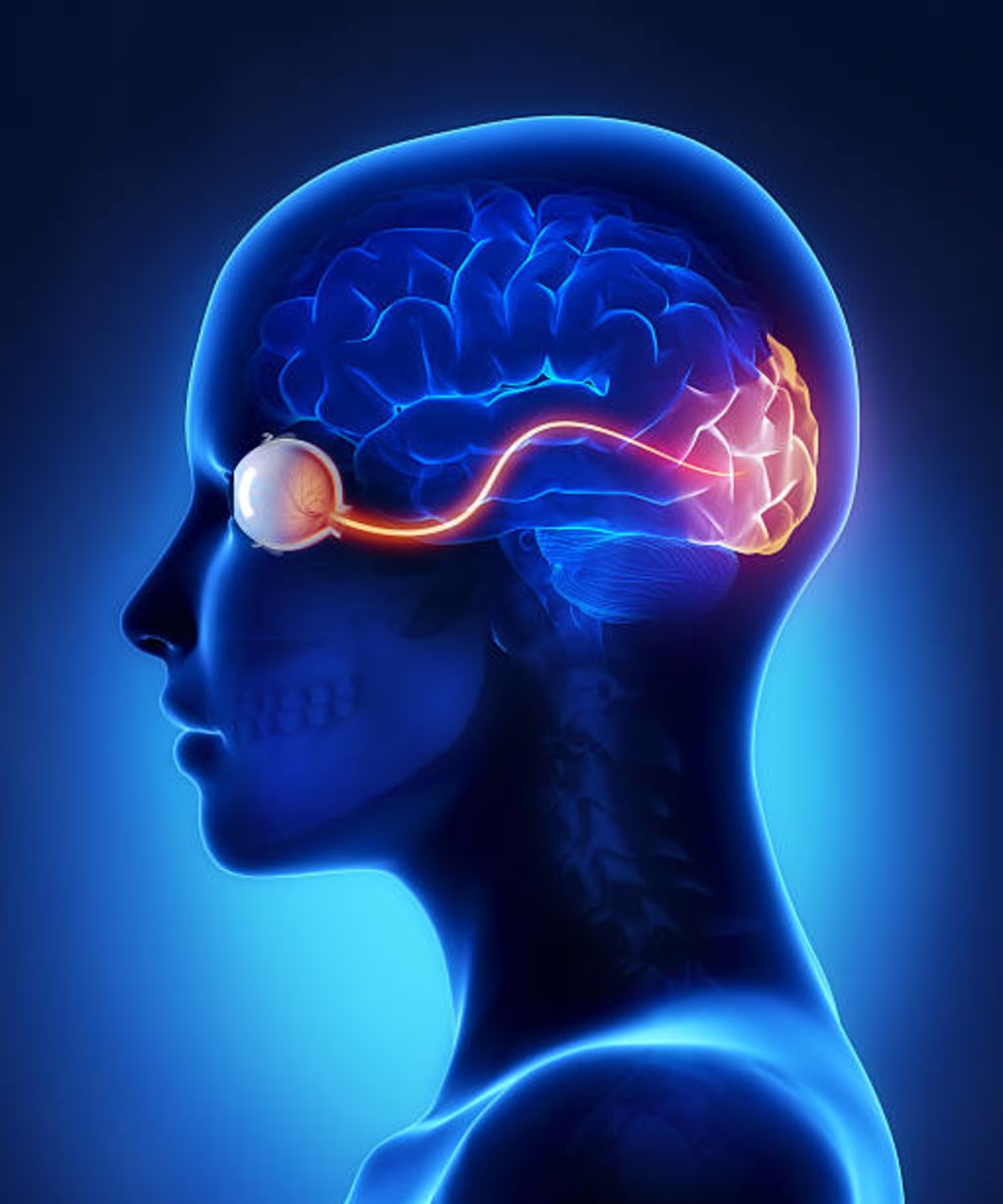
The Visual Pathway: From Retina to Brain:
The visual pathway involves the transmission of signals from the retina to the brain. Axons from retinal ganglion cells form the optic nerve, which passes through the lateral geniculate nuclei in the thalamus before reaching the primary visual cortex. The left visual field is processed in the right primary visual cortex, and vice versa.
Organization of the Visual Cortex:
The primary visual cortex, located in the occipital lobes, receives input from the lateral geniculate nuclei. Neurons in the primary visual cortex project to the secondary visual cortex and the visual association cortex. The secondary visual cortex receives input from the primary cortex, while the visual association cortex integrates visual information from multiple brain regions. The visual cortex is organized into vertical columns, corresponding to specific retinal areas.
Visual Analysis and Streams of Information:
Visual analysis occurs in the secondary visual cortex and the visual association cortex. Neurons in the dorsal stream interpret spatial information and motion, while neurons in the ventral stream process object characteristics, such as color and shape. These streams transmit information from the primary visual cortex to various regions in the brain, forming a hierarchy of visual processing.
Conclusion:
The journey of visual processing from the moment light enters the eye to the interpretation of visual information by the brain is a complex and remarkable process. Understanding the intricacies of the visual cortex and the mechanism of visual transduction provides a glimpse into the incredible capabilities of the human visual system. As we continue to explore the wonders of neuroscience, let us appreciate the intricate dance of light, neurons, and perception that unfolds within our visual cortex.
About the Creator
Keep reading
More stories from Nadula disanayaka and writers in Education and other communities.
The Cycle of Life and Death
Reincarnation, the belief in the rebirth of a soul after death, is a fundamental concept in many Eastern religions, including Buddhism. For Buddhists, reincarnation is intimately connected to the concept of karma and the understanding of the cyclical nature of existence. In this article, we will explore the intricate relationship between reincarnation and Buddhism, shedding light on its significance and implications within the Buddhist worldview.
By Nadula disanayaka11 months ago in Education
The Illumination of the Soul
In the heart of a bustling city, amidst the ceaseless rhythm of life, there dwelled a young woman named Maya. She navigated through the urban sprawl with a heavy heart, her days filled with monotony and a sense of longing for something more. Despite her outward composure, Maya yearned for a spark to ignite the depths of her soul, to breathe life into her existence.
By Labu Hossain7 days ago in Education


Comments
There are no comments for this story
Be the first to respond and start the conversation.