
"Sweet Victory: How to Crush Diabetes with the Right Treatment Plan and Mindset"
Diabetes Mellitus : A Comprehensive Overview

Diabetes Mellitus
An increase in plasma blood glucose is a clinical syndrome known as diabetes mellitus (hyperglycemia). Diabetes can have numerous origins, however type 1 or type 2 diabetes are the most frequent ones.
In contrast to type 2 diabetes, which is characterised by resistance to the action of insulin and an inability to create enough insulin to overcome this "insulin resistance," type 1 diabetes is brought on by the autoimmune destruction of insulin-producing cells ( cells) in the pancreas. Problems resulting from hyperglycemia might be immediate or long-lasting. Acutely, elevated blood sugar and insulin deficiency can cause noticeable symptoms, metabolic decompensation, and hospitalisation.
Diabetes-specific "microvascular" issues affecting the feet (nephropathy), kidneys (nephropathy), and eyes (retinopathy) are brought on by chronic hyperglycemia (neuropathy).
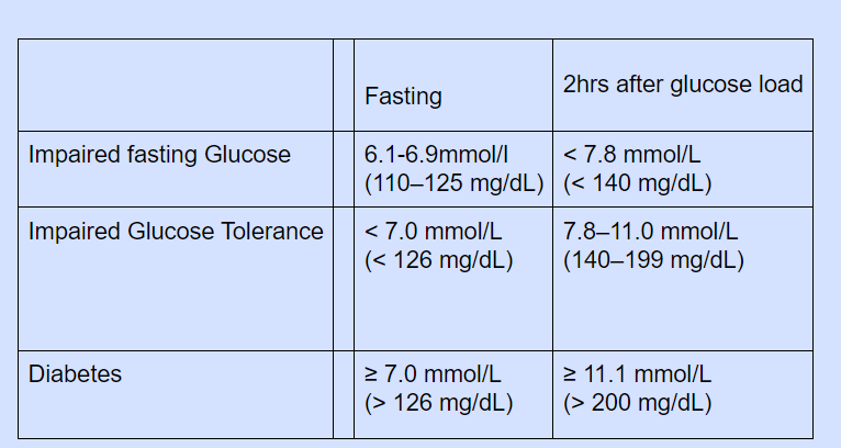
The population's blood glucose levels are distributed continuously, with no obvious distinction between those with normal and abnormal readings. The diagnostic criteria for diabetes (fasting plasma glucose 7.0 mmol/L (126 mg/dL) or glucose 11.1 mmol/L (200 mg/dL) 2 hours after an oral glucose challenge) have been chosen to identify those who have a level of hyperglycemia that, if untreated, carries a sizable risk of microvascular disease, particularly diabetic retinopathy. Impaired glucose tolerance is a term used to describe less severe hyperglycemia. This is linked to a higher risk of big vessel disease, such as atheroma leading to myocardial infarction, as well as a higher risk of acquiring diabetes in the future, although it is not significantly associated with a higher risk of microvascular illness.
Clinical Signs of a diabetic person:
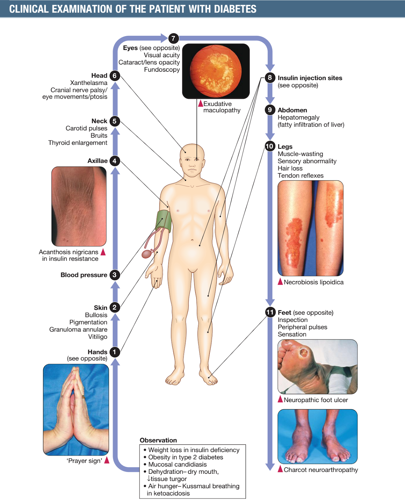
Population Affected:
People of all ages, genders, and ethnicities are affected by the global health issue known as diabetes. According to the World Diabetes Federation, there were 468 million persons worldwide who had diabetes in 2019, accounting for 9.3% of all adults. By 2045, this figure is anticipated to reach 700 million.
The prevalence of diabetes varies greatly by geographic location, with low- and middle-income nations having the greatest rates. In 2019, China (116 million cases), India (77 million cases), and the United States had the highest rates of adult diabetes (34 million).
The burden of diabetes is particularly high in certain populations, including Indigenous peoples, migrant populations, and those living in poverty. In many cases, these groups face significant barriers to accessing diabetes care, including limited access to healthcare services, medications, and education.
In addition to the human toll of diabetes, the condition also has significant economic costs. According to the International Diabetes Federation, global healthcare spending on diabetes was estimated at $760 billion in 2019. This includes both direct costs (such as healthcare services and medications) and indirect costs (such as lost productivity).
Given the significant impact of diabetes on global health and well-being, there is an urgent need for continued research and investment in diabetes prevention, treatment, and management. This includes efforts to improve access to healthcare services and medications, as well as initiatives to promote healthy lifestyles and prevent diabetes through education and awareness-raising campaigns.
Between meals, intestinal glucose absorption decreases, which causes a decrease in portal vein insulin and glucose concentrations and an increase in glucagon levels. As a result, gluconeogenesis and glycogen breakdown boost hepatic glucose output. Now that net glucose production has resumed, glucose homeostasis is sustained. Glycerol and amino acids are the primary substrates for gluconeogenesis.
FUNCTIONAL ANATOMY &
PHYSIOLOGY:
Normal glucose and fat metabolism:
The limited range of blood glucose is strictly controlled and maintained. For the central nervous system to receive a constant supply of glucose, this is crucial. The blood-brain barrier is mostly impermeable to fatty acids, and the brain has little capacity to store energy in the form of glycogen or triglycerides, thus it needs on the liver for a steady supply of glucose for oxidation and the subsequent production of adenosine triphosphate (ATP). The achievement of glucose homeostasis involves the coordinated actions of numerous organs, but it primarily reflects a balance between the uptake of glucose by peripheral tissues, especially skeletal muscle and the brain, and the entry of glucose into the circulation from the liver, supplemented by intestinal absorption of glucose after meals.
After ingestion of a meal containing carbohydrate, normal blood glucose levels are maintained by:
• suppression of hepatic glucose production
• stimulation of hepatic glucose uptake
• stimulation of glucose uptake by peripheral tissues
In response to an increase in blood glucose, pancreas cells produce insulin, the main regulator of glucose metabolism and storage, into the portal circulation. A number of other substances, including amino acids and hormones like glucagon-like peptide 1 (GLP-1) and gastrointestinal peptide, produced from the gut after eating can increase insulin secretion (GIP). Due to this phenomenon known as the "incretin effect," more insulin is released when glucose is given orally as opposed to when the same rise in plasma glucose is produced by intravenous glucose infusion.
In combination with a decrease in portal glucagon concentrations, the rise in portal vein insulin and glucose following a meal limits the liver's ability to produce glucose and leads to net hepatic glucose uptake. Around one-quarter to one-third of the glucose that is consumed is absorbed in the liver, depending on the amount of carbohydrates consumed. Furthermore, insulin increases the absorption of glucose in skeletal muscle and fat through the glucose transporter GLUT 4. Between meals, intestinal glucose absorption decreases, which causes a decrease in portal vein insulin and glucose concentrations and an increase in glucagon levels. As a result, gluconeogenesis and glycogen breakdown boost hepatic glucose output. Now that net glucose production has resumed, glucose homeostasis is sustained.
Glycerol and amino acids are the primary substrates for gluconeogenesis. Triglycerides are created by adipocytes (and the liver) from non-esterified (or "free" fatty acids) and glycerol. Not only is fatty acid metabolism heavily regulated by insulin, but also the metabolism of glucose. After meals, high insulin levels encourage lipid accumulation. In contrast, during a fast, low insulin levels allow for lipolysis and the release of FFAs (and glycerol) into the bloodstream, which can then be oxidised by a variety of organs. In addition to producing ketone bodies (acetoacetate, which can be reduced to 3-hydroxybutyrate or decarboxylated to acetone), their partial oxidation in the liver also provides energy to power gluconeogenesis. Organic acids called ketones are oxidised and used as metabolic fuel when they are generated in modest amounts.
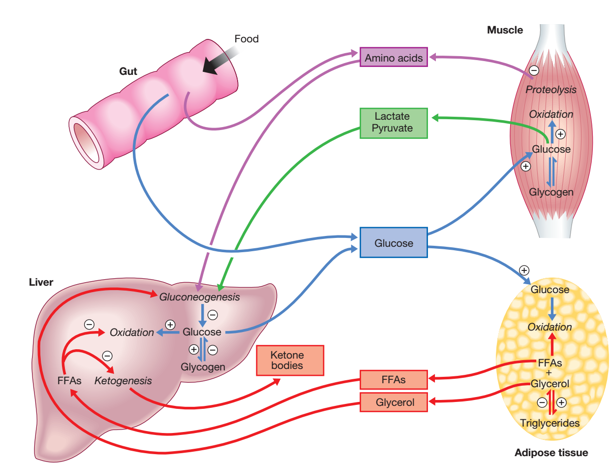
The pace at which ketone bodies may be utilised by peripheral tissues is constrained, and hyperketonemia develops when the liver produces more ketone bodies than it can remove. When insulin levels are low and catecholamine levels are high during famine, this physiological phenomenon takes place, increasing lipolysis and FFA transport to the liver.
Aetiology and pathogenesis of diabetes:
The exact causes of diabetes are not fully understood, but both genetic and environmental factors are believed to play a role. In type 1 diabetes, the immune system attacks and destroys the cells in the pancreas that produce insulin, whereas in type 2 diabetes, the body becomes resistant to insulin or doesn't produce enough of it to regulate blood glucose levels.
Environmental variables and genetic predisposition work together in both prevalent forms of diabetes to determine who gets the clinical syndrome and when it starts. Yet, type 1 and type 2 diabetes have very different underlying genes, triggering environmental events, and pathophysiology.
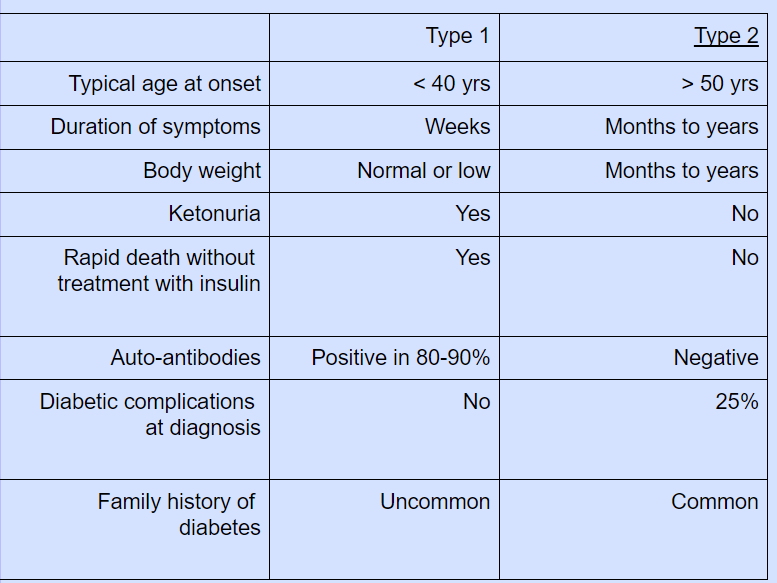
Insulin-dependent diabetes mellitus (IDDM), the old name for type 1 diabetes, is generally characterised by severe insulin shortage necessitating replacement medication. Because patients with type 2 diabetes still have some insulin secretion capacity but have impaired insulin sensitivity (insulin resistance), it was previously referred to as "non-insulin-dependent diabetes mellitus" (NIDDM). Initially, insulin replacement therapy is typically not necessary to treat type 2 diabetes.
Yet, 20% or more of type 2 diabetes patients will eventually experience severe insulin shortage necessitating replacement medication.
Type 1 diabetes:
Pathology:
The T cell-mediated autoimmune condition known as type 1 diabetes results in the death of the insulin-secreting cells in the pancreatic islets. Long-term (months to years) progressive loss of cell function occurs, but severe hyperglycemia and the hallmark diabetes symptoms don't appear until 80–90% of the cells' functional capacity has been reduced.
Insulitis, a pathology characterised by the invasion of the islets by mononuclear cells containing activated macrophages, helper cytotoxic and suppressor T lymphocytes, natural killer cells, and B lymphocytes, characterises the pre-diabetic pancreas pathology. These lesions begin as patchy lesions, and up until a very late stage, adjacent to unaffected lobules, lobules with strongly infiltrated islets are seen.
The destructive process is β cell specific, the glucagon and other hormone-secreting cells in the islet remaining intact. Islet cell antibodies are present prior to the clinical presentation of type 1 diabetes, and their identification can be helpful in establishing a diagnosis, but they are not very reliable indicators of how the illness will develop and eventually disappear. Type 1 diabetes is associated with other autoimmune disorders including thyroid disease, coeliac disease, Addison’s disease, pernicious anaemia and vitiligo.
Genetic predisposition:
About one-third of the polygenic inheritance of type 1 diabetes susceptibility is due to genetic factors. More than 20 different areas of the human genome have been linked to type 1 diabetes, but the IDDM 1 locus on the short arm of chromosome 6's major histocompatibility complex—which contains the human leucocyte antigen (HLA) region—has received the most attention. With the neighbouring alleles of the HLA-DQA1 and DQB1 genes, the HLA haplotypes DR3 and/or DR4 are in "linkage disequilibrium," which means they tend to be passed together and enhance the risk of type 1 diabetes in Caucasians. Since these HLA class II genes encode proteins on the surface of cells that deliver foreign and self antigens to T lymphocytes, they may be the primary genetic risk factors. Several genes, such as CD25, PTPN22, IL2RA, and IL-10, which are involved in immunological detection of pancreatic islet antigens, T-cell development, and immune modulation, have also been linked to type 1 diabetes through candidate gene and genome-wide association studies.The genes associated with type 1 diabetes overlap with those for other autoimmune disorders, such as coeliac disease and thyroid disease, consistent with clustering of these conditions in individuals or families.
Environmental predisposition:
Although genetic susceptibility appears to be a prerequisite for type 1 diabetes, the concordance rate between monozygotic twins is less than 40%, and wide geographic and seasonal variations in
incidences suggest that environmental factors have an important role in precipitating disease.Although hypotheses abound, the nature of these environmental factors are uncertain. They may trigger type 1 diabetes through direct toxicity to β cells or by
stimulating an autoimmune reaction directed against β cells. Potential candidates fall into three main categories:
viruses, specific drugs or chemicals, and dietary constituents. Viruses implicated in the aetiology of type 1 diabetes include mumps, Coxsackie B4, retroviruses, rubella (in utero), cytomegalovirus and Epstein–Barr virus. Various dietary nitrosamines (found in smoked and cured meats) and coffee have been proposed as potentially diabetogenic toxins. Bovine serum albumin (BSA), a major constituent of cow’s milk, has been implicated, since children who are given cow’s milk early in infants are more likely to develop type 1 diabetes than those who are breastfed. BSA may cross the neonatal gut and raise antibodies which cross-react with a heat-shock protein expressed by β cells. It has also been proposed that reduced exposure to microorganisms in early childhood limits maturation of the immune system and increases susceptibility to autoimmune disease (the ‘hygiene hypothesis’).
Metabolic disturbances in type 1 diabetes:
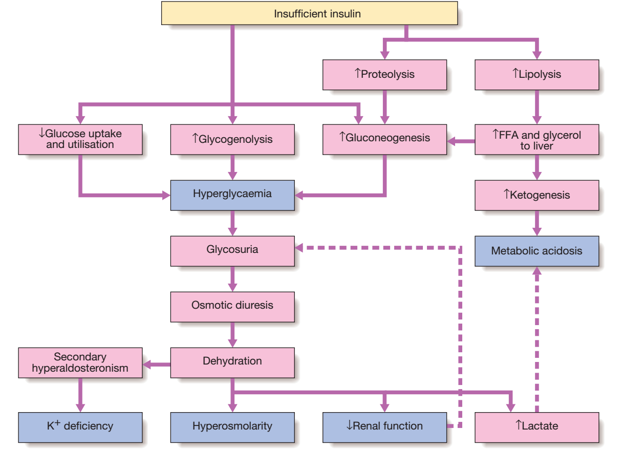
Patients with type 1 diabetes present when progressive β-cell destruction has crossed a threshold at which adequate insulin secretion and normal blood glucose levels can no longer be sustained.Above a certain level, high glucose levels may be toxic to the remaining β cells, so that profound insulin deficiency rapidly ensues, causing the metabolic sequelae.Hyperglycemia leads to glycosuria and dehydration, causing fatigue, polyuria, nocturia, thirst and polydipsia, susceptibility to urinary and genital tract infections, and later tachycardia and hypotension.Unrestrained lipolysis and proteolysis result in weight loss. Ketoacidosis occurs when generation of ketone bodies exceeds the capacity for their metabolism. Elevated blood H+ ions drive K+ out of the intracellular compartment, while secondary hyper-aldosteronism encourages urinary loss of K+. Thus patients usually present with a short history (typically a few weeks) of hyperglycaemic symptoms (thirst, polyuria, nocturia and fatigue), infections and weight loss, and may have developed ketoacidosis.
Type 2 diabetes:
Pathology:
Type 2 diabetes is a highly heterogeneous diagnosis that is made after type 1 diabetes and other types of diabetes have been ruled out.
In order to keep blood glucose levels within normal range, insulin resistance initially causes increased insulin secretion. The pancreatic cells, however, are unable to keep up with the increased demand for insulin in susceptible individuals, and an insulin shortage slowly worsens over time. Some patients, especially the elderly, develop diabetes despite not being obese and may have more pronounced -cell failure. These patients are typically driven by insulin resistance caused by obesity and ethnicity at a young age. The distinguishing characteristic is a "relative" insulin shortage, which means that not enough insulin is produced to overcome insulin action resistance.
In contrast, type 1 diabetes causes a rapid decline in insulin production and an utter lack of the hormone, leading to ketoacidosis and, if the insulin is not restored, death.
Insulin resistance:
The precursor to type 2 diabetes, poor glucose tolerance, is one of a number of illnesses that are thought to be brought on by resistance to insulin action. As a result, patients with type 2 diabetes frequently also have hypertension, dyslipidemia, non-alcoholic fatty liver, and, in women, polycystic ovarian syndrome. Dyslipidemia is characterised by elevated levels of small dense low-density lipoprotein (LDL) cholesterol and triglycerides and a low level of high-density lipoprotein (HDL) cholesterol.
The "insulin resistance syndrome" or "metabolic syndrome" is the name given to this group of symptoms, which is significantly more prevalent in individuals who are fat.
Although the underlying aetiology of insulin resistance is yet unknown, it is likely that several abnormalities in insulin signalling exist, impacting many tissues. One idea focuses on the adipocyte, which is particularly intriguing given that obesity is a significant factor in elevated insulin resistance. The metabolically active intra-abdominal "central" adipose tissue releases substantial amounts of FFAs, which may promote insulin resistance because they compete with glucose as a source of energy for oxidation in peripheral tissues like muscle.Moreover, adipose tissue secretes several hormones, including a range of peptides known as "adipokines" because they share structural similarities with immunological "cytokines," which act on particular receptors to affect the sensitivity of other tissues to insulin. Because visceral adipose tissue drains into the portal vein, central obesity may have a particularly strong effect on the liver's sensitivity to insulin, which would then have a negative impact on gluconeogenesis and hepatic lipid metabolism.
Another significant factor affecting insulin sensitivity is physical activity. Inactivity may encourage the buildup of FFAs in skeletal muscle and is linked to the down-regulation of insulin-sensitive kinases. Hence, those who are sedentary and have the same level of obesity are more insulin resistant than those who are active. Moreover, exercise enables glucose uptake into muscle that is not insulin-dependent, lowering the need for pancreatic cells to make insulin.
A typical symptom of central obesity is the accumulation of fat in the liver, which is made worse by insulin resistance and/or insufficiency. Non-alcoholic fatty liver disease (NAFLD) is a common complication of type 2 diabetes and is characterised by fatty infiltration of the liver. Although the condition may go better with effective diabetes and dyslipidemia treatment, some patients still develop non-alcoholic steatohepatitis and cirrhosis.
Pancreatic β-cell failure:
There is a small loss in the overall mass of pancreatic islet tissue in the early stages of type 2 diabetes. Around 50% of -cell function is lost at the time of diagnosis, and this decreases over time.
Type 2 diabetes is characterised by a few pathological alterations, the most recurrent of which is amyloid accumulation in the islets. Moreover, increased FFAs and plasma glucose have toxic effects on pancreatic cells that reduce insulin production. Although there are fewer -cells, their mass is unaltered, and glucagon secretion is enhanced. This may result in hyperglycemia.
Genetic predisposition:
Genetic factors play an important role in type 2 diabetes, as shown by the marked differences in susceptibility among different ethnic groups and studies of identical twins, in which concordance rates for type 2 diabetes are nearly 100%. However, many genes are involved, and the likelihood of developing diabetes is also strongly influenced by environmental factors.
Genome-wide association studies have identified over 65 genes or gene regions associated with type 2 diabetes, with each gene having a small effect. The largest effect is seen with variations in TCF7L2; the 10% of the population with two copies of the risk variant for this gene have an almost twofold increased risk of developing type 2 diabetes. Most genes known to contribute to the risk of type 2 diabetes are involved in β-cell function or cell cycle regulation and cell turnover, suggesting that altered regulation of β-cell mass is a key factor.
Environmental and other risk factors:
Diet and obesity: Epidemiological studies show that type 2 diabetes is associated with excessive diet, especially when combined with obesity and lack of exercise. Middle-aged people with diabetes eat significantly more, are fatter and less active than their non-diabetic siblings. The risk of developing type 2 diabetes increases tenfold in people with a body mass index (BMI) of more than 30 kg/m2. Although the majority of patients with type 2 diabetes are obese, only a minority of obese people develop diabetes because the majority of obese patients are able to increase insulin secretion to compensate for the increased demand due to obesity and insulin resistance. Those who develop diabetes may have genetically impaired β-cell function, reduced β-cell mass or susceptibility of β-cells to attack by toxic substances such as FFAs or inflammatory cytokines
Age:
1.Prevalence: increases with age, affecting ~10% of people over 65 years. Half of these people are undiagnosed.
Impaired β-cell function and exaggerated insulin resistance with ageing both contribute.
2.Glycosuria: the renal threshold for glucose rises with age, so glycosuria may not develop until the blood glucose concentration is markedly raised.
3.Pancreatic carcinoma: may present in old age with the development of diabetes, in association with weight loss and diminished appetite.
Forms of diabetes:
1)Type 1 diabetes:
• Immune-mediated
• Idiopathic
2)Type 2 diabetes.
3) Other specific types:
• Genetic defects of β-cell function.
• Genetic defects of insulin action (e.g. leprechaunism,lipodystrophies)
• Pancreatic disease (e.g. pancreatitis, pancreatectomy,neoplastic disease, cystic fibrosis, haemochromatosis,fibrocalculous pancreatopathy)
• Excess endogenous production of hormonal antagonists to insulin
Growth hormone – acromegaly,
Glucocorticoids – Cushing’s syndrome,
Glucagon – glucagonoma,
Catecholamines – pheochromocytoma,
Thyroid hormones – thyrotoxicosis
•Drug-induced (e.g. corticosteroids, thiazide diuretics,
phenytoin)
•Uncommon forms of immune-mediated diabetes (e.g. IPEX (immunodysregulation polyendocrinopathy X) syndrome)
• Associated with genetic syndromes (e.g. Down’s syndrome;Klinefelter’s syndrome; Turner’s syndrome;(Wolfram’s syndrome) – diabetes insipidus, diabetes mellitus,optic atrophy, nerve deafness; Friedreich’s ataxia; myotonic dystrophy)
4)Gestational diabetes.
INVESTIGATIONS:
Urine analysis:
Glucose:
Urine glucose testing with test sticks is a common screening procedure for diabetes. If possible, the test should be performed on urine given 1-2 hours after a meal to maximise sensitivity. Glycosuria always warrants further assessment by blood test. The main disadvantage of measuring glucose in urine is the individual variation in the kidneys' threshold for glucose. The most common cause of glycosuria is a low kidney threshold, which is common in pregnancy and in young people; the resulting 'renal glycosuria' is a benign condition unrelated to diabetes. Another drawback is that some drugs (such as β-lactam antibiotics, levodopa and salicylates) can interfere with urine glucose tests.
Ketone bodies:
Ketone bodies can be detected by the nitroprusside reaction, which measures acetoacetate, using tablets or dipsticks. Ketonuria can be found in normal people who have fasted or exercised strenuously for long periods, who have vomited repeatedly, or who have eaten a high-fat, low carbohydrate diet. Ketonuria is therefore not pathognomonic for diabetes, but if it is accompanied by glycosuria, the diagnosis of diabetes is very likely. In diabetic ketoacidosis, ketones can also be detected in the plasma with test sticks.
Protein:
The standard dipstick test for albumin detects urine albumin in concentrations greater than 300 mg/L. Smaller amounts can only be measured with special albumin dipsticks or by quantitative biochemical laboratory measurements.
Microalbuminuria or proteinuria is an important indicator of diabetic nephropathy and/or increased risk of macrovascular disease in the absence of urinary tract infection.
Blood testing:
Glucose:
Laboratory tests of blood glucose levels are based on an enzymatic reaction (glucose oxidase) and are cheap, usually automated and very reliable. However, the blood glucose level depends on whether the patient has eaten anything recently. Therefore, it is important to consider the circumstances under which the blood sample was taken. Blood glucose can also be measured with colorimetric or other test sticks, which are often read with a portable electronic metre. These are used for capillary (finger prick) tests to monitor diabetes management. It is controversial whether self-monitoring improves blood glucose control in people with type 2 diabetes.
In many countries, self-monitoring is now only offered to people with type 2 diabetes who are receiving insulin therapy.
To diagnose diabetes, blood glucose concentrations should be determined using an accurate laboratory method rather than a portable technique.
The glucose concentration is lower in venous blood than in arterial or capillary blood (finger prick). The glucose concentration in whole blood is lower than that in plasma because red blood cells contain relatively little glucose. Venous plasma values are usually the most reliable for diagnostic purposes
Ketones:
Monitoring of ketone levels in the blood is increasingly available.The urinary ketone measurements described above are semi-quantitative, difficult to perform, and retrospective (i.e. the urine has accumulated over several hours) and does not measure the most important ketone in the blood in diabetic ketoacidosis (DKA), beta-hydroxybutyrate (β-OHB). Whole blood ketone monitoring detects β-OHB and is useful to assist insulin adjustment in intercurrent illness or persistent hyperglycemia to prevent or detect DKA. Blood ketone monitoring is also useful to monitor the resolution of DKA in hospitalised patients.
Glycosylated haemoglobin:
Glycosylated haemoglobin is an accurate and objective measure of blood glucose control over a period of weeks to months.
In diabetes, the slow non-enzymatic covalent binding of glucose to haemoglobin (glycation) increases the proportion in the HbA1 (HbA1c) fraction relative to non-glycated haemoglobin (HbA0) in adults.
These fractions can be separated by chromatography; laboratories may report glycated haemoglobin as total glycated haemoglobin (GHb), HbA1 or HbA1c.
In most countries, HbA1c is the preferred measurement. The rate of formation ofHbA1c is directly proportional to the ambient blood glucose concentration; a 1% increase in HbA1c corresponds to an approximate average increase of 2 mmol/L (36 mg/dL) of blood glucose. Although HbA1c concentration reflects integrated glycaemic control over the life of the red cell (120 days), the HbA1c value is most sensitive to changes in glycaemic control that occur in the month prior to measurement.
Different test methods are used to measure HbA1c, but most laboratories report HbA1c values(in %) that follow the reference range used in the Diabetes Control and Complications Trial (DCCT).
To enable worldwide comparisons of HbA1c values, the International Federation for Clinical Chemistry and International Federation of Clinical Chemistry and Laboratory Medicine (IFCC) has developed a standard method; IFCC-standardised HbA1c values are expressed in mmol/mol. In 2011, many countries adopted the IFCC reference method.
HbA1c estimates may be falsely decreased in anaemia or during pregnancy and may be difficult to interpret in patients with uraemia or haemoglobinopathy with some test methods.
Establishing the diagnosis of diabetes:
Blood glucose levels i.e Hyperglycemia can be divided into three categories: normal, impaired (pre-diabetes) and diabetes. The threshold for diabetes is based on the level above which there is a significant risk of microvascular complications (retinopathy, nephropathy, neuropathy). People who are classified as prediabetic have blood glucose levels that carry a negligible risk of microvascular complications, but an increased risk of developing diabetes. Since the risk of macrovascular disease (atheroma in the large blood vessels) increases steadily as blood glucose levels rise in the population, people with prediabetes also have an increased risk of cardiovascular disease (heart attack, stroke and peripheral vascular disease).
Diabetes is confirmed by either:
•Plasma glucose in random sample or 2 hrs after a 75 g
glucose load ≥ 11.1 (200 mg/dL) or
•Fasting plasma glucose ≥ 7.0 mmol/L (126 mg/dL)
In asymptomatic patients, two diagnostic tests are required to
confirm diabetes.
“Pre-diabetes” is classified as:
•Impaired fasting glucose = fasting plasma glucose ≥ 6.0
(108 mg/dL) and < 7.0 mmol/L (126 mg/dL)
•Impaired glucose tolerance = fasting plasma glucose
< 7.0 mmol/L (126 mg/dL) and 2-hr glucose after 75 g oral
glucose drink 7.8–11.1 mmol/L (140–200 mg/dL).
Oral glucose tolerance test (OGTT):
Which patients to test:
• Fasting plasma glucose 6.1–7.0 mmol/L (110–126 mg/dL).
• Uncertainty about the diagnosis of diabetes.
Preparation before the test:
• Unrestricted carbohydrate diet for 3 days.
• Fasted overnight for at least 8 hrs.
• Rest for 30 mins.
• Remain seated for the duration of the test, with no smoking.
Sampling:
• Measure plasma glucose before and 2 hrs after a 75 g oral
glucose drink.
Interpretation (venous plasma glucose):
Symptoms of hyperglycemia:
. Thirst, dry mouth
• Polyuria
• Nocturia
• Tiredness, fatigue, lethargy
• Change in weight (usually weight loss)
• Blurring of vision
• Pruritus vulvae, balanitis (genital candidiasis)
• Nausea
• Headache
• Hyperphagia; predilection for sweet foods
• Mood change, irritability, difficulty in concentrating, apathy.
Onset of Diabetes:
Diabetic ketoacidosis (DKA):
Diabetic ketoacidosis (DKA) is a serious complication of diabetes that occurs when the body begins to break down fat instead of glucose (sugar) for energy due to a lack of insulin. This process leads to an accumulation of acidic by-products called ketones in the blood, resulting in acidosis.
DKA is most common in people with type 1 diabetes, but can also occur in people with type 2 diabetes, especially those who are overweight or obese. DKA is usually triggered by a combination of factors, such as an illness or infection, inadequate insulin dosage or stress.
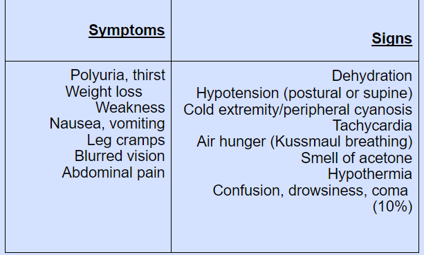
Symptoms of DKA may include excessive thirst, frequent urination, nausea, vomiting, abdominal pain, shortness of breath, confusion and fruity-smelling breath. If left untreated, DKA can progress to a life-threatening condition such as coma or death. Treatment for DKA usually involves hospitalisation and administration of intravenous fluids, insulin and electrolytes to correct the imbalance caused by ketones. It is important for people with diabetes to monitor their blood glucose levels regularly and see a doctor immediately if they experience symptoms of DKA.
A clear understanding of the biochemical basis and pathophysiology of DKA is essential for its efficient treatment. The cardinal biochemical features are:
• hyperketonaemia (≥ 3 mmol/L) & ketonuria (more than 2+ on standard urine sticks)
• hyperglycaemia (blood glucose ≥ 11 mmol/L(~200 mg/dL))
• metabolic acidosis (venous bicarbonate<15 mmol/L and/or venous pH < 7.3)
Management of diabetic ketoacidosis:
DKA is a medical emergency which should be treated in hospital, preferably in a high-dependency area. If available, the diabetes specialist team should be involved. Regular clinical and biochemical review is essential, particularly during the first 24 hours of treatment.
Insulin:
A fixed-rate intravenous insulin infusion of 0.1 U/kg body weight/hr is recommended.
Exceptionally, if intravenous administration is not feasible, soluble insulin can be given by intramuscular injection (loading dose of 10–20 U, followed by 5U-hourly), or a fast-acting insulin analogue can be given hourly by subcutaneous injection (initially 0.3 U/kg body weight, then 0.1 U/kg hourly). The blood glucose concentration should fall by 3–6 mmol/L (approximately 55–110 mg/dL) per hour, or blood ketone concentrations fall by at least 0.5 mmol/L/hr.
A more rapid decrease in blood glucose should be avoided, as this might precipitate hypoglycaemia and the serious complication of cerebral oedema, particularly in children. Failure of blood glucose to fall within 1 hour of commencing insulin infusion should lead to a re-assessment of insulin dose. Ketosis, dehydration, acidaemia, infection and stress combine to produce severe insulin resistance in some cases, but most will respond to a low-dose insulin regimen. When the blood glucose has fallen, 10% dextrose infusion is introduced and insulin infusion continues to encourage glucose uptake into cells and restoration of normal metabolism. In recent years, it has also become increasingly common to continue with the use of long-acting insulin analogues administered subcutaneously during the initial management of DKA; this provides background insulin for when the intravenous insulin is discontinued.Restoration of the usual insulin regimen, by subcutaneous injection, should not be instituted until the patient is both biochemically stable and able to eat and drink normally.
Fluid replacement:
In adults, rapid fluid replacement in the first few hours is usually recommended. Caution is recommended in children and young adults because of the risk of cerebral oedema. Most current guidelines favour correction of the extracellular fluid deficit with isotonic saline (0.9% sodium chloride). If the plasma sodium is greater than 155 mmol/L, 0.45% saline may be used initially.
Potassium:
Careful monitoring of potassium is essential to the management of diabetic ketoacidosis because both hypo and hyperkalaemia can occur and are potentially life-threatening. Potassium replacement is not usually recommended with the initial litre of fluid because prerenal failure may be present secondary to dehydration.
Treatment with 0.9% sodium chloride with potassium chloride 40 mmol/L is recommended if the serum potassium is below 5.5 mmol/L and the patient is passing urine. If the potassium falls below 3.5 mmol/L, the potassium replacement regimen needs to be reviewed. Cardiac rhythm should be monitored in severe DKA because of the risk of electrolyte-induced cardiac arrhythmia.
Bicarbonate:
Adequate fluid and insulin replacement should resolve the acidosis. The use of intravenous bicarbonate therapy is currently not recommended. Acidosis may reflect an adaptive response, improving oxygen delivery to the tissues, and so excessive bicarbonate may induce a paradoxical increase in cerebrospinal fluid acidosis and has been implicated in the pathogenesis of cerebral oedema in children and young adults.
Hyperglycaemic hyperosmolar state(HHS):
Hyperglycaemic hyperosmolar state (HHS) is characterised by severe hyperglycaemia (> 30 mmol/L (600 mg/dL)), hyperosmolality (serum osmolality > 320 mOsm/kg), and dehydration in the absence of significant hyperketonaemia (< 3 mmol/L) or acidosis (pH > 7.3, bicarbonate > 15 mmol/L). It was previously referred to as hyperosmolar non-ketotic (HONK) coma but, as in DKA, coma is not invariable. As with DKA, there is glycosuria, leading to an osmotic diuresis, with loss of water, sodium, potassium and other electrolytes.
However, in HHS, hyperglycaemia usually develops over a longer period (a few days to weeks), causing more profound hyperglycaemia and dehydration (fluid loss may be 10–22 litres in a person weighing 100 kg). The reason that patients with HHS do not develop significant ketoacidosis is unclear, although it has been speculated that insulin levels may be too low to stimulate
glucose uptake in insulin-sensitive tissues, but still sufficient to prevent lipolysis and subsequent ketogenesis.
A mixed picture of HHS and DKA can occur. Although typically occurring in the elderly, HHS is increasingly seen in younger adults. Common precipitating factors include infection, myocardial infarction, cerebrovascular events or drug therapy (e.g. corticosteroids). Poor prognostic signs include hypothermia, hypotension (systolic blood pressure < 90 mmHg), tachy- or bradycardia, severe hypernatraemia (sodium > 160 mmol/L), serum osmolality > 360 mOsm/kg, and the presence of other serious comorbidities. Mortality rates are higher than in DKA – up to 20% in the USA, reflecting the age and frailty of the population and more frequent presence of comorbidities.
The aims are to normalise osmolality, replace fluid and electrolyte losses, and normalise blood glucose, at the same time preventing complications such as arterial or venous thrombosis, cerebral oedema and central pontine de-myelinosis. Comorbidities also need to be taken into account; for example, rapid fluid replacement may precipitate cardiac failure in patients with coronary artery disease. Historically, management of HHS has followed DKA guidelines, but increasing recognition of the differences between HHS and DKA has led to new approaches
in HHS. In particular, rapid shifts in osmolality should be avoided through more measured fluid replacement regimens that are guided by serial calculations of serum osmolality. A key recommendation is that 0.9% sodium chloride solution alone is used for initial treatment, and that insulin is introduced only when the rate of fall in blood glucose has plateaued.
The normal value is 280–290 mmol/L and consciousness is impaired when it is high (> 340 mmol/L), as commonly occurs in HHS.
Hypoglycaemia:
Hypoglycaemia (blood glucose < 3.5 mmol/L (63 mg/dL)) in diabetes results in most circumstances from insulin therapy, less frequently from use of oral insulin secretagogues such as sulphonylurea drugs, and rarely with other anti-diabetic drugs. When hypoglycaemia develops in non-diabetic people, it is called ‘spontaneous’ hypoglycaemia.
Hypoglycaemia can be a frequent occurrence in the lives of people with type 1 diabetes and has a major impact on their willingness and ability to achieve target glucose levels.In health, a number of mechanisms are in place to ensure that glucose homeostasis is maintained. If blood glucose falls, three primary physiological defence mechanisms operate: endogenous insulin release from pancreatic β cells is suppressed; release of glucagon from pancreatic α cells is increased; and the autonomic nervous system is activated, with release of catecholamines both systemically and within the tissues. In addition, stress hormones, such as cortisol and growth hormone, are increased in the blood. These actions reduce whole-body glucose uptake and increase hepatic glucose production, maintaining a glucose supply to the brain. People with type 1 diabetes cannot regulate insulin once it is injected subcutaneously, and so it continues to act, despite developing hypoglycaemia. In addition, within 5 years of diagnosis, most patients will have lost their ability to release glucagon specifically during hypoglycaemia. This is thought to result mainly from loss of α-cell regulation by β cells. These two primary defects mean that hypoglycaemia occurs much more frequently in people with type 1 and longer duration type 2 diabetes.
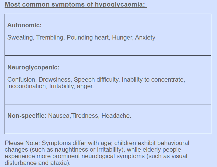
Causes of hypoglycaemia:
Missed, delayed or inadequate meal.
Unexpected or unusual exercise.
Alcohol.
Errors in oral anti-diabetic agent(s) or insulin dose/schedule/administration.
Poorly designed insulin regimen particularly if predisposing to nocturnal hyper-insulinaemia.
Lipohypertrophy at injection sites causing variable insulin absorption.
Gastroparesis due to autonomic neuropathy causing variable carbohydrate absorption.
Malabsorption, e.g. coeliac disease.
Unrecognised other endocrine disorder, e.g. Addison’s disease
Factitious (deliberately induced).
Breastfeeding.
Risk factors for severe hypoglycaemia:
Strict glycaemic control
Impaired awareness of hypoglycaemia
Age (very young and elderly)
Long duration of diabetes
Sleep
C-peptide negativity (indicating complete insulin deficiency)
History of previous severe hypoglycaemia
Renal impairment
Genetic, e.g. angiotensin-converting enzyme (ACE) genotype
Management:
Treatment of hypoglycaemia depends on its severity and on whether the patient is conscious and able to swallow. Oral carbohydrate usually suffices if hypoglycaemia is recognised early. If parenteral therapy is required, then as soon as the patient is able to swallow, glucose should be given orally.
Full recovery may not occur immediately and reversal of cognitive impairment may not be complete until 60 minutes after normoglycaemia is restored. When hypoglycaemia has occurred in a patient treated with a long- or intermediate-acting insulin or a long-acting sulphonylurea, such as glibenclamide, the possibility of recurrence should be anticipated; to prevent this, infusion of 10% dextrose, titrated to the patient’s blood glucose, may be necessary.
If the patient fails to regain consciousness after blood glucose is restored to normal, then cerebral oedema and other causes of impaired consciousness – such as alcohol intoxication, a post-ictal state or cerebral haemorrhage – should be considered. Cerebral oedema has a high mortality and morbidity, and requires urgent treatment with mannitol and high-dose oxygen.
Following recovery, it is important to try to identify a cause and make appropriate adjustments to the patient’s therapy. Unless the reason for a hypoglycaemic episode is clear, the patient should reduce the next dose of insulin by 10–20% and seek medical advice about further adjustments in dose.
Prevention of hypoglycaemia:
Patient education is fundamental to the prevention of hypoglycaemia. Risk factors for, and treatment of hypoglycaemia should be discussed. The importance of regular blood glucose monitoring and the need to have glucose (and glucagon) readily available should be stressed. A review of insulin and carbohydrate management during exercise is particularly useful.
Avoidance and treatment of hypoglycaemia:
Carry a supply of fast-acting carbohydrate (non-perishable, in suitable containers) Screw-top plastic bottles for glucose drinks Packets of powdered glucose (for use in hot, humid climates), Confectionery (foil-wrapped in hot climates)
Companions should carry additional oral carbohydrate, and glucagon
Perform frequent blood glucose testing (carry spare metre and/or visually read strips)
Use fast-acting insulin analogues for long-distance air travel.
Diabetes in pregnancy:
During pregnancy, maternal glucose metabolism changes to optimise glucose and other nutrient delivery to the fetus. This is particularly apparent in the second half of pregnancy, when there is an increase in maternal tissue insulin resistance, such that glucose is preferentially supplied to the fetus rather than maternal tissue.
This is largely driven by the maternal hormonal environment, with increased oestrogens and progestogens, and,
in particular, human placental lactogen (hPL). The delivery of the placenta results in rapid decline in hPL with a rapid reversal of insulin resistance soon after birth.
During pregnancy, fasting plasma glucose decreases slightly, while post-prandial blood glucose may be increased. The renal threshold for glycosuria is reduced in pregnancy.
In the fetus, insulin secretion is driven by fetal blood glucose levels, which are determined by the maternal glucose concentrations. Thus maternal hyperglycaemia drives fetal hyperinsulinaemia. Since insulin is a major fetal growth factor, hyperinsulinaemia in turn drives increased fetal growth, resulting in increased birth weight (‘macrosomia’)
Gestational diabetes:
Gestational diabetes is defined as diabetes with first onset or recognition during pregnancy. This definition will include a few patients who develop type 1 diabetes during pregnancy, where prompt action and early insulin treatment will be required, and some patients who develop type 2 diabetes, or had unknown pre-existing type 2 diabetes, in whom the diabetes does not remit after pregnancy. However, the majority of gestational diabetes develops due to an inability to increase insulin secretion adequately to compensate for pregnancy-induced insulin resistance, and most women can expect to return to normal glucose tolerance immediately after pregnancy.
In contrast to non-gestational diabetes, for which the diagnostic thresholds for diabetes are based upon risk of microvascular complications, the diagnosis of gestational diabetes is based upon maternal blood glucose measures that are associated with increased fetal growth. An international consensus recommended that glucose values diagnostic of gestational diabetes should be lower than those for non-gestational diabetes. Controversy remains about who should be screened, and in part the screening strategy depends on the population risk. It is widely accepted that women at high risk for gestational diabetes should have an oral glucose tolerance test at 24–28 weeks, with some guidelines recommending that all are screened by measuring HbA1c, fasting blood glucose or random blood glucose at the first booking visit. With the increasing use of HbA1c to diagnose diabetes, it should be noted that HbA1c is unreliable after early pregnancy, when it falls due to increased red cell turnover.
Identifying patients with gestational diabetes:
Women at high risk of gestational diabetes
•BMI > 30 kg/m2
• A previous macrosomic baby weighing ≥ 4.5 kg at birth
•Previous gestational diabetes
• A first-degree relative with diabetes
•A high-risk ethnicity – South Asian, black Caribbean or Middle Eastern
Diagnosis:
•High-risk women should have a 75 g oral glucose tolerance test before 28 weeks’ gestation.
•Gestational diabetes is diagnosed when:
Fasting plasma glucose ≥ 5.1 mmol/L (92 mg/dL) or 1-hour plasma glucose (after glucose load) ≥ 10 mmol/L (180 mg/dL) or 2-hour plasma glucose (after glucose load) ≥ 8 mmol/L (144 mg/dL).
•Consider testing high-risk women at first booking visit with an HbA1c or fasting blood glucose.
Management of gestational diabetes:
The aim is to normalise the maternal blood glucose and thereby reduce excessive fetal growth. The first element of management is dietary modification, in particular by reducing consumption of quick-acting refined carbohydrates. Women with gestational diabetes should undertake regular pre- and post-prandial self-monitoring of blood glucose, aiming for pre-meal blood glucose levels of less than 5.5 mmol/L (100 mg/dL) or post-meal blood glucose levels of less than 7.0 mmol/L (125 mg/dL). If treatment is necessary, metformin or glibenclamide is considered safe to use in pregnancy.
Glibenclamide should be used rather than other sulfonylureas because it does not cross the placenta. Other oral therapies or injectable incretin-based therapies should not be given in pregnancy. Insulin may be required, especially in the later stages of pregnancy. If the maternal blood glucose is not well controlled prior to, and during, delivery, the resulting fetal hyperinsulinemia leads to neonatal hyper-insulinaemia, which in turn can cause neonatal hypoglycaemia.
After delivery, maternal glucose usually rapidly returns to pre-pregnancy levels. Women should be tested at least 6 weeks postpartum with an oral glucose tolerance test. In women with persistent mild fasting
hyperglycaemia (glucose over 5.5 mmol/L (100 mg/dL)) who have a small increment (less than 3.5 mmol/L) in plasma glucose 2 hours after oral glucose, monogenic diabetes due to a mutation in GCK (encoding glucokinase) should be considered.
Those who have returned to normal glucose tolerance remain at considerable risk for developing type 2 diabetes, with a 5-year risk between 15 and 50%, depending on the population. Therefore, all women who have had gestational diabetes should be given diet and lifestyle advice to reduce their risk of developing type 2 diabetes.
Diabetes in adolescence:
Type of diabetes: type 1 diabetes is predominant in children and adolescents, but type 2 diabetes is now presenting in unprecedented numbers of obese, inactive teenagers. Monogenic diabetes (MODY) should also be considered
Physiological changes: hormonal, physical and lifestyle changes in puberty affect dietary intake, exercise patterns and sensitivity to insulin, necessitating alterations in insulin regimen.
Emotional changes: adolescence is a phase of transition into independence (principally from parental care).Periods of rebellion against parental control, experimentation (e.g. with alcohol) and a more chaotic lifestyle are common, and often impact adversely on control of diabetes.
Glycaemic control: a temporary deterioration in control is common, although not universal. It is sometimes more important to maintain contact and engagement with a young person than to insist on tight glycaemic control.
Diabetic ketoacidosis: a few adolescents and young adults present with frequent episodes of DKA, often because of non-adherence to insulin therapy. This is more common in females. Motivating factors may include weight loss, rebellion, and manipulation of family or schooling circumstances.
Adolescent diabetes clinics: these challenges are best tackled with support from a specialised multidisciplinary team, including paediatricians, physicians, nurses and psychologists. Support is required for the patient and parents.
Complications of diabetes:
Microvascular/neuropathic:
Retinopathy, cataract
•Impaired vision
Nephropathy
•Renal failure
Peripheral neuropathy
•Sensory loss
• Pain
•Motor weakness
Autonomic neuropathy
•Gastrointestinal problems (gastroparesis; altered bowel habit)
• Postural hypotension
Foot disease
• Ulceration
•Arthropathy
Macrovascular:
Coronary circulation
•Myocardial ischaemia/infarction
Cerebral circulation
•Transient ischaemic attack
•Stroke
Peripheral circulation
•Claudication
•Ischaemia
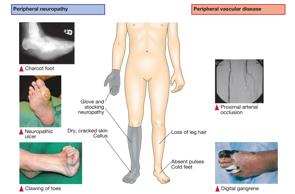
Diabetic neuropathy :
Diabetic Neuropathy is a type of nerve damage that can occur as a result of diabetes. High levels of blood sugar can damage nerves throughout the body, leading to a variety of symptoms. Here are some key aspects to consider:
Types of diabetic neuropathy: There are several types of diabetic neuropathy, including:
Peripheral neuropathy: This is the most common type of diabetic neuropathy and affects the nerves in the feet and legs, although it can also affect the hands and arms. Symptoms can include numbness, tingling, burning, or sharp pain.
Autonomic neuropathy: This type of neuropathy affects the nerves that control involuntary functions, such as digestion, heart rate, and blood pressure. Symptoms can include bladder problems, gastrointestinal issues, and sexual dysfunction.
Proximal neuropathy: This type of neuropathy affects the nerves in the hips, thighs, and buttocks. Symptoms can include pain, weakness, and difficulty standing up from a seated position.
Focal neuropathy: This type of neuropathy affects a specific nerve or group of nerves and can cause sudden weakness or pain in a specific area.
Risk factors: The risk of developing diabetic neuropathy increases with the duration of diabetes, as well as poor blood sugar control. Other risk factors can include high blood pressure, high cholesterol, smoking, and obesity.
Diagnosis: Diabetic neuropathy is typically diagnosed based on symptoms and a physical exam. Additional tests may be recommended, such as nerve conduction studies or electromyography, to assess the extent of nerve damage.
Treatment: Treatment for diabetic neuropathy typically focuses on managing symptoms and preventing further nerve damage. This can include:
Blood sugar control: Keeping blood sugar levels within a target range can help prevent further nerve damage.
Medications: Medications such as antidepressants, anticonvulsants, and opioids may be used to manage pain associated with diabetic neuropathy.
Lifestyle changes: Quitting smoking, maintaining a healthy weight, and exercising regularly can help improve blood sugar control and reduce the risk of further nerve damage.
Foot care: People with diabetic neuropathy should pay close attention to foot care, including daily inspections for cuts or sores, wearing comfortable shoes, and avoiding activities that could cause foot injuries.
Diabetic retinopathy:
Diabetic retinopathy is a condition in which high blood sugar levels damage blood vessels in the retina, the light-sensitive tissue at the back of the eye. Here are some key aspects to consider:
Types of diabetic retinopathy: There are two main types of diabetic retinopathy:
Nonproliferative diabetic retinopathy: This is the early stage of diabetic retinopathy and occurs when small blood vessels in the retina become damaged and leak fluid or blood into the retina. This can cause swelling and blurred vision.
Proliferative diabetic retinopathy: This is the more advanced stage of diabetic retinopathy and occurs when new blood vessels grow on the surface of the retina. These new vessels are fragile and can bleed into the eye, leading to severe vision loss or even blindness.
Risk factors: The risk of developing diabetic retinopathy increases with the duration of diabetes, as well as poor blood sugar control. Other risk factors can include high blood pressure, high cholesterol, smoking, and pregnancy.
Symptoms: In the early stages, diabetic retinopathy may not cause any noticeable symptoms. As the condition progresses, symptoms can include:
Blurred or distorted vision
Floaters or spots in the field of vision
Loss of vision or dark spots in the center of the visual field
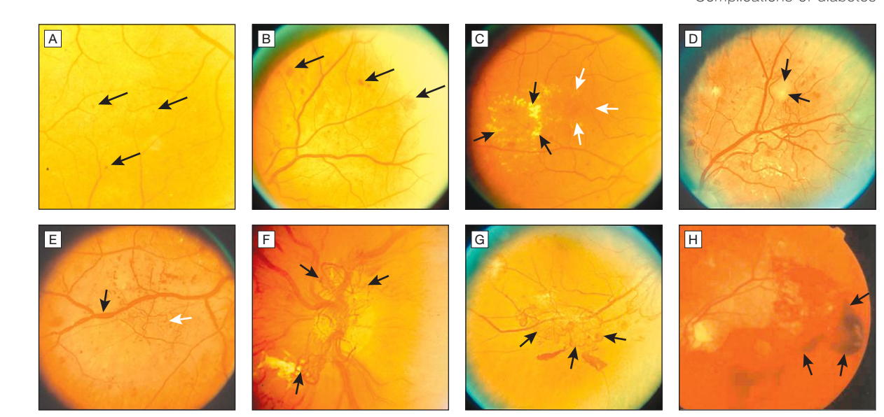
Diagnosis: Diabetic retinopathy is typically diagnosed through a comprehensive eye exam, which can include visual acuity testing, dilation of the pupils, and examination of the retina. Additional tests, such as optical coherence tomography (OCT) or fluorescein angiography, may be recommended to assess the extent of retinal damage.
Treatment: Treatment for diabetic retinopathy depends on the severity of the condition. In the early stages, close monitoring and management of blood sugar levels may be all that is needed.
As the condition progresses, treatment options may include:
Laser treatment: Laser therapy can be used to seal leaking blood vessels or reduce the growth of abnormal blood vessels.
Intravitreal injections: Medications can be injected directly into the eye to reduce inflammation or prevent the growth of abnormal blood vessels.
Vitrectomy: In severe cases, surgery may be necessary to remove blood and scar tissue from the eye.
It is important for people with diabetes to have regular eye exams to monitor for diabetic retinopathy and other eye conditions. Early detection and treatment can help prevent vision loss and maintain good eye health.
Management of Diabetes:
Medication:
There are several types of medications used in the management of diabetes, including:
Insulin: Insulin is a hormone produced by the pancreas that helps regulate blood sugar levels. In people with type 1 diabetes, the pancreas produces little or no insulin, so insulin therapy is necessary. In people with type 2 diabetes, insulin may also be used if other medications are not effective in controlling blood sugar levels. Insulin can be administered through injections or an insulin pump.
Oral medications: There are several classes of oral medications that can be used to manage type 2 diabetes, including:
Metformin: This medication helps reduce the amount of glucose produced by the liver and improves the body's ability to use insulin.
Sulfonylureas: These medications stimulate the pancreas to produce more insulin.
DPP-4 inhibitors: These medications help reduce blood sugar levels by increasing the production of insulin and decreasing the amount of glucose produced by the liver.
GLP-1 receptor agonists: These medications help increase insulin production and decrease glucose production by the liver. They also slow down the rate at which food leaves the stomach, which can help reduce blood sugar spikes after meals.
SGLT2 inhibitors: These medications help reduce blood sugar levels by increasing the amount of glucose excreted in the urine.
Injectable medications: In addition to insulin, there are several injectable medications that can be used to manage type 2 diabetes, including:
GLP-1 receptor agonists: These medications, such as exenatide and liraglutide, can be given once or twice daily by injection. They work by stimulating insulin production and decreasing glucose production by the liver.
Amylin mimetics: These medications, such as pramlintide, work by slowing down the rate at which food leaves the stomach, which can help reduce blood sugar spikes after meals.
Diet: Eating a healthy and balanced diet is important for managing diabetes. This includes limiting processed and sugary foods, choosing whole grains, lean proteins, and healthy fats, and eating a variety of fruits and vegetables. It is also important to monitor carbohydrate intake and spread out meals and snacks throughout the day.
Exercise: Regular physical activity can help improve blood sugar control and reduce the risk of complications associated with diabetes. This can include a variety of activities such as walking, biking, swimming, or strength training. Aim for at least 30 minutes of moderate-intensity exercise most days of the week.
Monitoring blood sugar: Checking blood sugar levels regularly is important for diabetes management. This can help identify patterns and make adjustments to medications, diet, and activity levels as needed. It is important to work with a healthcare provider to determine the target range for blood sugar levels.
Managing stress: Stress can affect blood sugar levels, so finding ways to manage stress is important for diabetes management. This can include techniques such as deep breathing, meditation, or yoga.
Quitting smoking: Smoking can increase the risk of complications associated with diabetes, so quitting smoking is important for overall health and diabetes management.
Regular check-ups: Regular check-ups with a healthcare provider are important for monitoring blood sugar levels, adjusting medications as needed, and identifying and managing any complications associated with diabetes
About the Creator
Saraj Rajkant
A medical Professional, a vocalist and a so so guitarist.. Learning, sharing and creating
Enjoyed the story? Support the Creator.
Subscribe for free to receive all their stories in your feed.
Comments
There are no comments for this story
Be the first to respond and start the conversation.