
Content warning
This story may contain sensitive material or discuss topics that some readers may find distressing. Reader discretion is advised. The views and opinions expressed in this story are those of the author and do not necessarily reflect the official policy or position of Vocal.
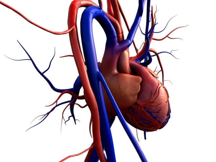
Blood Flow through the Heart
Blood flowing procedure and related diseases

The heart is an organ that pumps blood throughout the body.
It has four main parts: two upper chambers called atria and two lower chambers called ventricles.
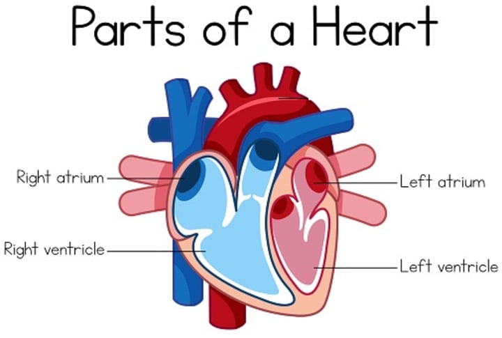
The Atria receive blood from the body and lungs, and the Ventricles pump blood to the lungs and body. The heart is divided into right and left sides by a wall of tissue.
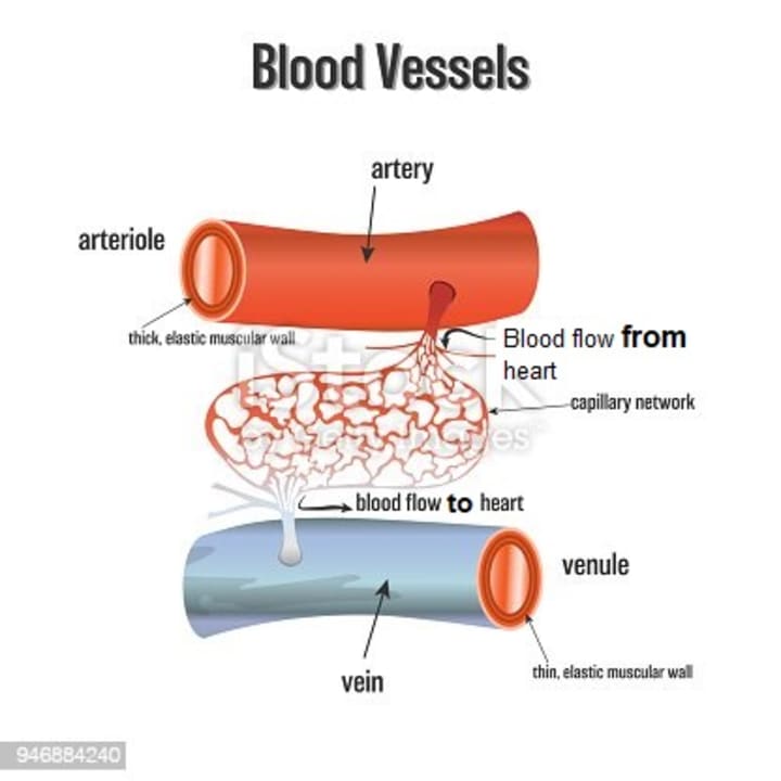
Veins: are blood vessels containing blood flowing to the heart.
Arteries: are blood vessels containing blood flowing from the heart.
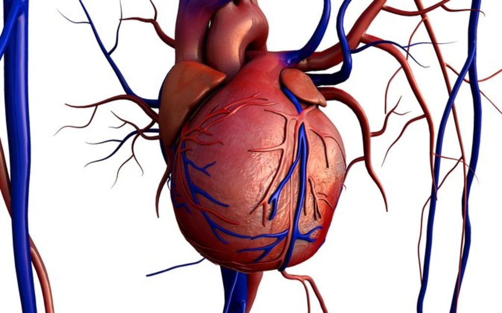
The blue is representative of blood vessels containing deoxygenated blood, while the red blood vessels have oxygenated blood fresh from the lungs. Now, let's follow the path of the blood through the heart.
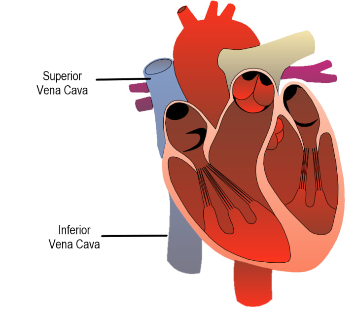
The superior vena-cava receives blood from the head, neck, upper limbs and chest. Meanwhile, the inferior vena-cava receives blood from the trunk, viscera, and lower limbs. both superior and inferior vena-cava end up in the right atrium, one of the four chambers of the heart. The heart not only has four chambers, it also has four valves. The purpose of the valves is to keep blood moving in the right direction and not flow backwards. Blood exits the right atrium through the tricuspid valve so called because it has three flaps, and enters the right ventricle.
The blood exits the right ventricle through the pulmonary valve and enters the pulmonary artery. Again, it is an artery because blood is flowing away from the heart, but it is blue because it lacks oxygen. The pulmonary artery then splits into the left and right pulmonary arteries, which go to each respective lung.
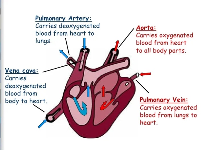
In the lungs, gas exchange occurs. The blood discards carbon dioxide and picks up oxygen. Now, blood comes back from the lungs through the pulmonary veins, entering the left atrium.
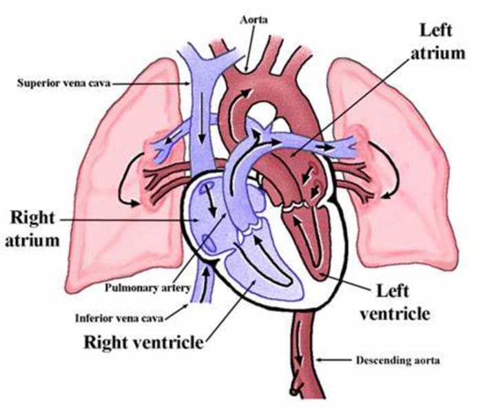
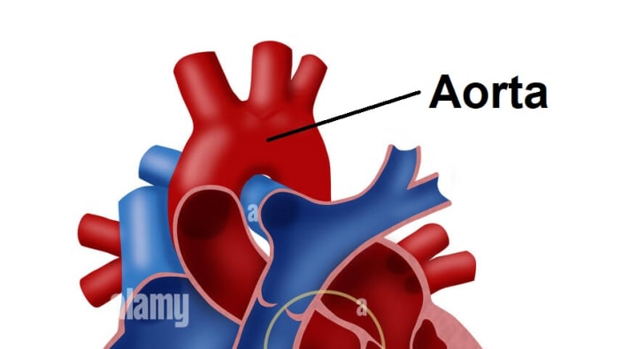
Next, the blood is pumped into the left into the left ventricle through the mitral, or bicuspid valve. finally, the oxygenated blood leaves the left ventricle through the aortic semi-lunar valve, entering the aortic arch.
The Aorta, which is the largest of all the arteries, distributes the oxygenated blood to the rest of the body. The aortic arch has three major branches, which supply the head and arms with blood.
Then, the aorta curls downward from behind the heart, forming the descending aorta, which descends through the chest and continues down through the abdomen. In the abdomen, the descending aorta splits to supply the pelvis and legs with blood.
HEART DISEASES
There are several heart disorders that can be caused by problems with the flow of blood through the heart. Here are a few common ones:
Aortic Stenosis: This is a condition where the aortic valve in the heart doesn't open fully, which restricts blood flow from the left ventricle to the aorta. It can cause symptoms like chest pain, fatigue, and fainting.
Mitral Valve Prolapse: In this condition, the mitral valve doesn't close properly, allowing some blood to flow backward into the left atrium. It often doesn't cause symptoms but can lead to chest pain or palpitations.
Atrial Septal Defect (ASD): ASD is a congenital heart defect where there's a hole in the wall (septum) that separates the two upper chambers of the heart (atria). This hole allows oxygen-rich blood to mix with oxygen-poor blood, affecting the overall blood flow in the heart.
Ventricular Septal Defect (VSD): VSD is another congenital heart defect, but this one involves a hole in the septum between the two lower chambers of the heart (ventricles). It can lead to oxygen-rich and oxygen-poor blood mixing, causing various symptoms.
Patent Ductus Arteriosus (PDA): In a fetus, the ductus arteriosus is a blood vessel that allows blood to bypass the lungs. In some cases, it doesn't close properly after birth, leading to abnormal blood flow between the aorta and pulmonary artery.
Coarctation of the Aorta: This is a congenital narrowing of the aorta, which can obstruct blood flow from the heart to the rest of the body. It can lead to high blood pressure and other cardiovascular problems.
Tetralogy of Fallot: This is a congenital heart defect comprising four abnormalities in the structure of the heart. It leads to a mix of oxygen-rich and oxygen-poor blood and can cause symptoms like cyanosis (bluish skin) and poor growth.
Hypertrophic Cardiomyopathy: In this condition, the heart muscle becomes abnormally thick, which can obstruct blood flow out of the heart. It can lead to chest pain, shortness of breath, and even sudden cardiac arrest.
Eisenmenger Syndrome: This is a condition that can develop when a congenital heart defect, such as a VSD or ASD, causes irreversible pulmonary hypertension and abnormal blood flow. It can result in severe heart and lung problems.
These are just a few examples of heart disorders related to blood flow. It's important to note that the treatment and management of these conditions vary, and they can range from medications to surgical interventions, depending on the severity and specific diagnosis.
*If you suspect you or someone you know may have a heart disorder, it's crucial to seek medical evaluation and guidance from a healthcare professional.
About the Creator
Keep reading
More stories from Eiman Asif and writers in Education and other communities.
sorrowful facts
Someone told me: "Anger never exists, we do it ourselves." As described Anger never existed on its own, it's a feeling that a person emphasizes to feel himself, we create our anger through our perceptions, interpretations, and responses to events or circumstances.
By Eiman Asif3 months ago in Writers
Time for #2
This is the second article on my review of the ideas in The Four Agreements. Here's the first article: I knew that I'd move forward with this series, but I put it off as Agreement #2 is a big stumbling block for me. I'd like to say I decided to get out of my own way and thus wrote this, but that isn't the truth. (Since Agreement #1 is to Be Impeccable with Your Word I surely need to own up to that)
By Judey Kalchik 4 days ago in BookClub


Comments (1)
This article is fantastic—I appreciate its well-crafted and informative nature.