
Human Reproductive System
General Information on Reproductive System.
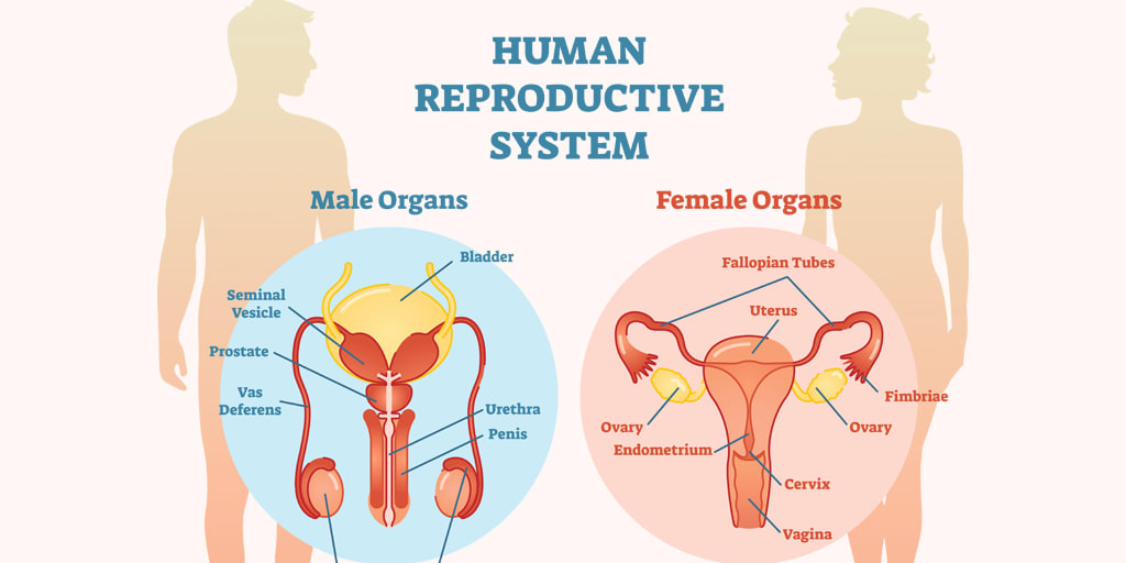
Reproductive system of human
It is a natural phenomenon by which organism reproduce young ones of their own kinds for continuation of race. Human beings are unisexual or dioecious i.e. male and female reproductive system is separated in separate individual.
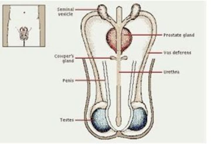
Male reproductive organs
1.Testes (one pair)
2.Epididymis (one pair)
3.Vasa deferens (one pair)
4.Ejaculatory duct (one pair)
5.Urethra.
6.Penis.
4.Accessory glands.
1.Testes: These are pinkish-oval lie inside the scrotum. The scrotum lies outside the abdominal cavity. So that temperature of testes remains 2.30c below than temperature of body which is needed to produces sperm.
Microscopic structure:
There are three layers to cover testis. These are Outer-tunica vaginalis, Middle-tunica albugania and Inner-layer-tunica vasculosa.

There are 200-300 testicular lobules in testis. Each lobule is composed of 1 to 4 coiled seminiferous tubules. The tubules are lined with germinal epithelial cells (spermatogonia).These epithelial cells produce sperms by the process of spermatogenesis. Sertoli cells supply nourishment to develop sperms.
There are interstitial cells (cells of leydig) in between seminiferous tubules and connective tissue. The interstitial cells produce androgens, which promotes development of male accessory glands and controls male sex features (moustache, beard, change of voice)
2.Epididymis:
It is funnel like convoluted about 6 meters long tube. Board part of anterior epididymis is called caput epididymis. Middle narrow part is corpus epididymis posterior end is cauda epididymis.
Function:
Store the sperm and secrets fluid to nourish sperm.
3.Vasa deferens:
It arises from cauda. It forms a loop around urinary bladder and joins with duct of seminal vesicle. It form ejaculatory duct with seminal vesicle.
Function:
carry sperm from testis to seminal vesicles.
4.Ejaculatory duct:
There are two short formed by the union of duct from a seminal vesicle and vasa deferens, there carry the mixture of sperm and secretion of seminal vesicle.
5.Urethra:
It is 20 cm long arises from urinary bladder. It discharges urine and semen both.
6.Penis:
It is copulatory organ of human. It is made up of three columns of spongy tissues (two dorsal cavernosa and one ventral corpora spongiosum). Enlarged-tip of penis is called glans penis which is highly sensitive. Penis deposits the semen in vagina with spermatozoa.
Accessory glands:
It consists of;
a.Seminal vesicles:
These are two pouches like below-posteriorly of urinary bladder and opens into ejaculatory duct. These secret and expel a viscous fluid which keep sperms alive.
b. Prostate gland:
It lies behind urinary bladder. It is secretes thin milky substance occupy about 30% volume of semen.
c.Cowper’s glands:
The paired glands lie below the prostate glands. Their alkaline secretion lubricates the semen flow.
d.Eretion of penis:
Erection is caused by dilation of blood vessel of blood vessel resulting collection of more blood spaces of spongy tissue. About 3ml of semen is discharged by penis in each ejaculation during copulation.
Female reproductive organs:
Female reproductive organs’ consists of following;
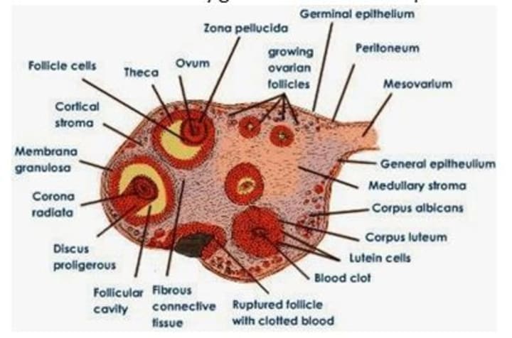
1.Overies:
There is one pair of about 3.5cm long, pink, almond shaped and situated in abdominal cavity on either side of vertebral column. Each kidney is attached with ovarian ligament and body wall with mesovarian fold with ovaries. Internally each ovary is differentiate with
i.Outer germinal epithelium (cubical)
ii.Tunica albunginea: connective tissue below germinal epithelium.
iii.Stroma: Inner mass of connective tissues made up of cortex medulla.
When Graafian follicle liberates ovum it is called corpus luteum. The corpus luteum secrete progesterone hormone in the influence of luteinizing hormone. The hormones thicken the endometrium wall and activate the mammary gland for their development.
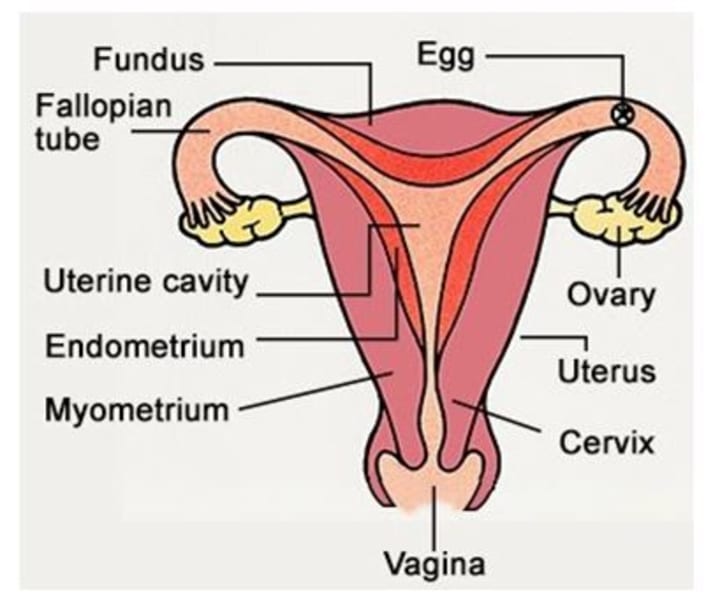
2.Fallopian tubes (oviducts): these are ciliated tubes of 10-20cm long arise from uterus.
It has four parts:
a.Funnel like infundibulum near ovary with finger like structure fimbriae
b.Wide curved part ampulla.
c.Narrow part isthmus.
d.Part near uterus (uterine).
3.Uterus: it is a pear shaped hollow muscular organ. Upper dome shaped part is called fundus. Main part is called body of uterus. Lower portion of uterus is called cervix.
4.Vagina: It is female copulatory organ. It is tubular, about 10cm long. It passage for menstrual flow and receptor of spermatozoa. In virgins the vaginal orifice is partially covered by hymen membrane.
5.Vulva: It is external genitalia it is consists of mons pubis (hair part) clitoris (erectile organ) labia major a (inner fold of vulva). Libia minora have in more number.
6.Accessory glands:
These consists of Bartholin’s glands and mammary glands.
Bartholin’s glands:
These are two bean-shaped lie on either of side of vaginal orifice These glands secret viscid fluid for lubricant of vulva during sexual intercourse and sexual excitement.
Mammary glands:
These are pair rounded with median nipple. Each gland made up of 15-25 lobules of milky glands. Each gland sends a lactiferous duct toward nipple. Milk gland produce milk under the control of prolactin and ejection of milk is controlled by pituitary gland.
About the Creator
Sumesh Bhaila
The main purpose of my writing is to motivate you people to do something that can help you achieve your big goals and dreams whatever they may be...
Please like & share it and also support me by leaving a tip.
Keep reading
More stories from Sumesh Bhaila and writers in FYI and other communities.
APJ Abdul Kalam
Dr. APJ Abdul Kalam is a world famous name. He is listed as one of the greatest scientists of the 21st century. In addition, he becomes the 11th president of India and has served his country. He was the most respected man in the country for his contribution as a scientist and as an incomparable President. Besides, his contribution to the ISRO (Indian Space Research Organization) is amazing. He has led many community projects and has been instrumental in the construction of Agni and Prithvi arrows. Through his involvement in Nuclear power in India, he was known as the “Missile Man of India”. And because of his contribution to the country, the government rewarded him with the highest public award.
By Sumesh Bhaila3 years ago in FYI
The Bloody Blood in Your Veins
Our Origins Today, I read something I didn't know—one more thing to add to all the other things I didn't know. By the time I grow up, I’ll have a collection. But it was intriguing, and it inspired me to write this post. I hope you enjoy it.
By Rene Volpi 5 days ago in FYI
Thank you Vocal team for a job well done
A year-long annoyance In January 2023, I began noticing that it took longer for my wallet page to load, sometimes up to five minutes. By January 2024, I could no longer see my wallet or request my funds. I tried from my cell phone to no avail, so I signed in from a different laptop. Everything worked, and I gave thanks, but the next time I tried, it did not work.
By Cheryl E Preston8 days ago in FYI


Comments
There are no comments for this story
Be the first to respond and start the conversation.