
MRI Coils: The Critical Hardware Most Patients Never Think About
The radiofrequency coil placed against your body during an MRI determines what doctors can and cannot see.

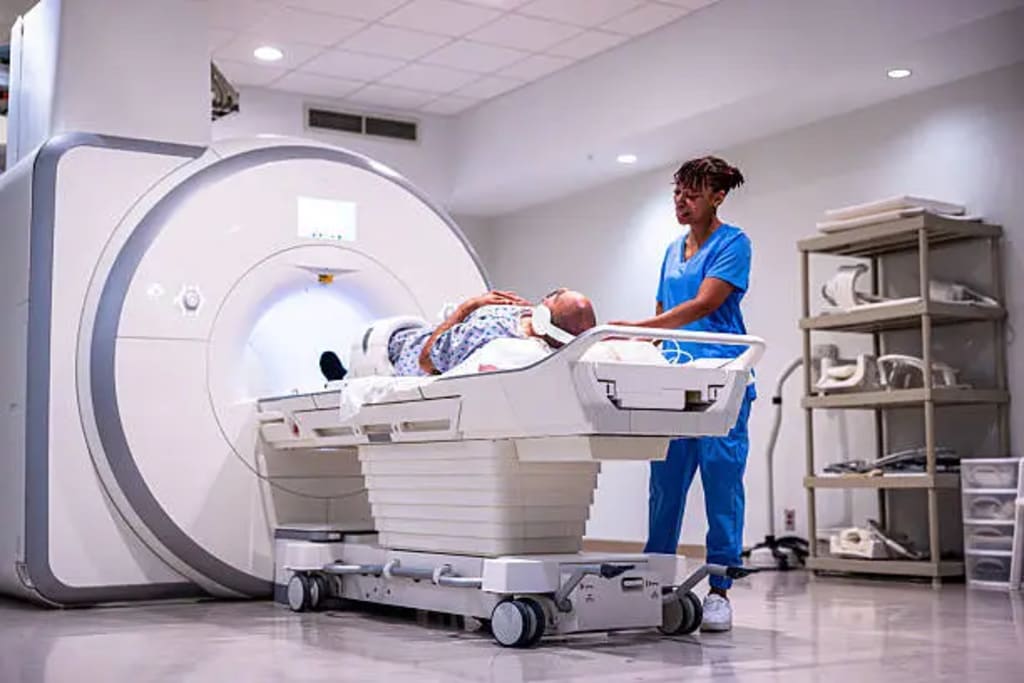
The Unsung Hardware Inside Every MRI Scanner That Determines What Doctors Actually See
Most people who have had an MRI remember the noise, the claustrophobia, and the instructions not to move. Very few remember — or were ever told about — the coil. Before the scan began, a technologist placed a piece of hardware against or around the body part being imaged: a close-fitting array for the knee, a cage-like structure around the head, a flat paddle beneath the spine. That hardware is an RF coil — a radiofrequency receiver that detects the signals emitted by hydrogen protons in the tissue being imaged and determines, more than almost any other single factor, the spatial resolution and signal-to-noise ratio of the resulting image.
The magnetic resonance imaging coils market is a specialized but technically critical segment of the broader medical imaging supply chain, and a comprehensive picture of its development is available in this MRI hardware and radiofrequency coil technology market analysis. The physics matter enormously here, and understanding them helps explain why this is a market with real commercial complexity.
Why Proximity Is Everything in MRI Signal Detection
MRI signal strength drops off with the square of the distance between the tissue being imaged and the detector — the coil. A coil positioned close to the anatomy of interest captures substantially more signal than the scanner's large body coil, which is built into the bore and surrounds the entire patient. More signal means better image quality, faster scan times, or both.
This physical reality drives the entire surface coil market. Dedicated coils for the brain, the shoulder, the wrist, the knee, the breast, the spine, the prostate — each designed to maximize signal capture from a specific anatomical region — are necessary clinical tools for diagnostic imaging. A knee MRI performed with a dedicated knee coil produces dramatically better images than one performed with a generic body coil, at a fraction of the scan time.
The Phased Array Revolution and What Came After
Modern clinical MRI coils are phased arrays — multiple small coil elements arranged in geometric patterns that work together, with their signals combined digitally to produce images with better coverage and signal characteristics than any single element could achieve. The introduction of phased array coils in the early 1990s was one of the most significant technical advances in clinical MRI, enabling parallel imaging techniques that accelerated scan times by factors of two to four without sacrificing diagnostic quality.
The evolution continues. Higher element counts — 32-channel, 64-channel, and even 128-channel arrays for specific applications like cardiac and neurological imaging — have become available, pushing image quality further. The technical challenge of managing signals from that many elements simultaneously has been addressed through advances in receiver electronics and digital signal processing that have largely happened inside the scanner itself.
The Market Structure and Who Controls It
The magnetic resonance imaging coils market operates in a somewhat peculiar competitive structure. The major MRI scanner manufacturers — Siemens Healthineers, GE HealthCare, and Philips — produce proprietary coils optimized for their own scanner platforms. These coils are not interchangeable across platforms; the interface between coil and scanner is proprietary, locking hospitals into single-manufacturer coil ecosystems for the life of their scanner.
This creates a classic razor-and-blade dynamic. Scanners are capital equipment with long replacement cycles. Coils need replacement or upgrading more frequently — through wear, technological obsolescence, or the need to support new imaging protocols. An independent coil manufacturer, Noras MRI Products, has worked to develop multi-platform compatible coils, but navigating the proprietary interface ecosystem has constrained the independent market's growth.
The Flexible Coil Frontier
One of the more interesting emerging segments in MRI coil technology involves flexible coil designs that conform to patient anatomy rather than presenting a rigid fixed geometry. Conventional coils are optimized for average anatomy and may not sit optimally against patients who are pediatric, obese, or have unusual anatomical features. Flexible coils — using thin-film conductor technologies or stretchable electronics — can conform more closely to the patient surface and potentially improve signal capture across a broader patient population.
The clinical and commercial implications of flexible coil technology extend beyond image quality into patient comfort and workflow. A coil that drapes naturally rather than requiring careful rigid positioning reduces technologist setup time and may improve the consistency of image quality across different operators and patient types.
About the Creator
Marvin M. Gibsonv
Marketing enthusiast and Digital Marketing Executive sharing practical tips on SEO, content creation, social media marketing, and online growth. Learn, grow, and stay ahead of digital trends.
Enjoyed the story? Support the Creator.
Subscribe for free to receive all their stories in your feed.
Comments
There are no comments for this story
Be the first to respond and start the conversation.