
AI in Microscopy: Seeing the Invisible and Speeding Up Drug Discovery
Deep learning is transforming what scientists can extract from microscope images — in labs and clinical pathology.

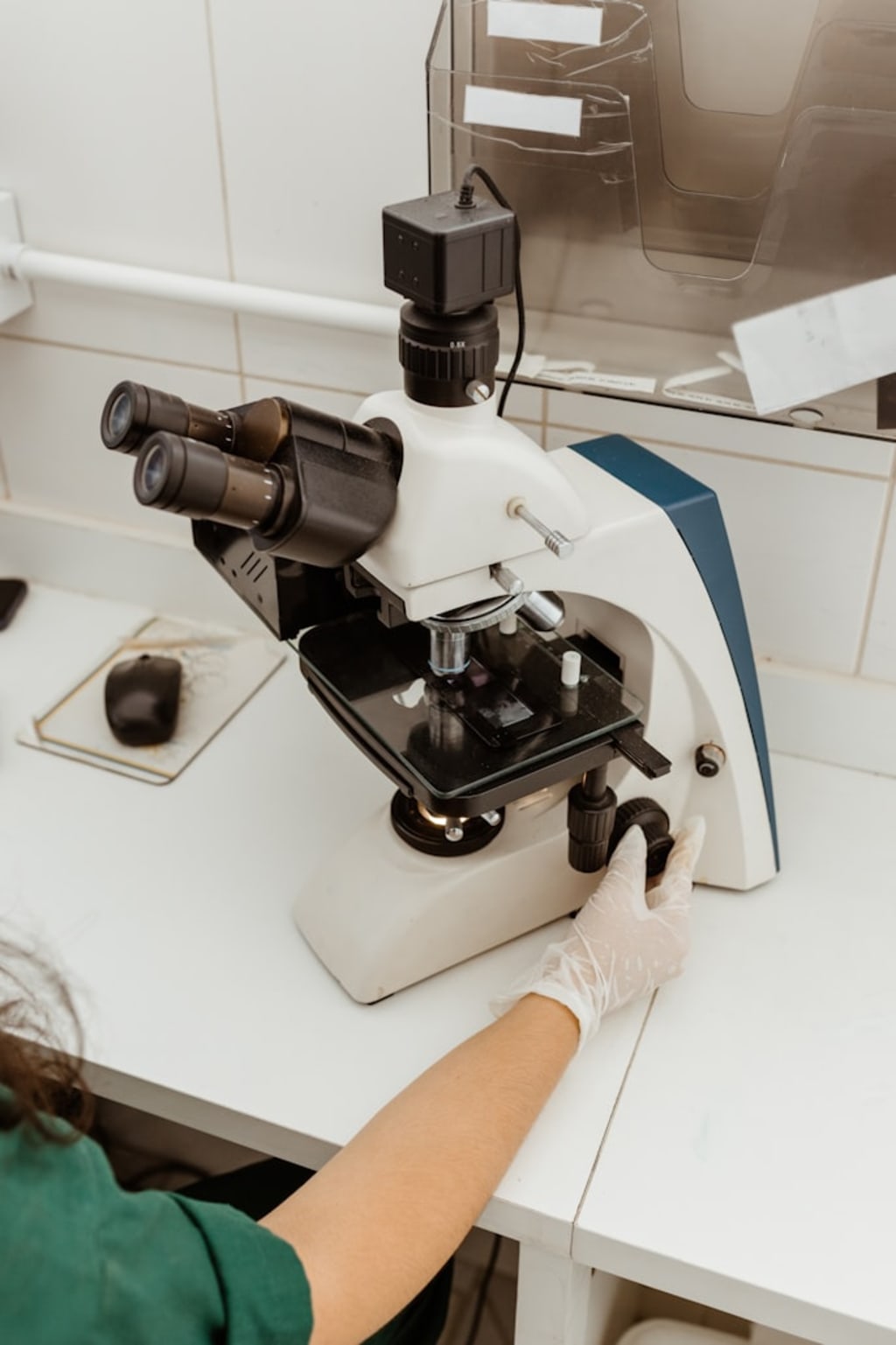
Scientists Can Now See Things Under a Microscope That Were Invisible Six Years Ago
In 2018, Google researchers published a paper describing a technique called CARE — Content-Aware Image Restoration — that used deep learning to reconstruct high-resolution microscopy images from low-dose, noisy acquisitions. The practical implication was startling: scientists could image living cells with less light exposure — reducing phototoxicity and photobleaching, two persistent problems in live cell imaging — and then have AI reconstruct the detail that the lower light conditions would otherwise have obscured. They were, in a meaningful sense, seeing more by imaging less.
This is the promise that has driven serious investment into AI in microscopy across academic research, pharmaceutical development, and clinical pathology. A broader analysis of where the AI in microscopy market is heading is available in this computational imaging and AI microscopy technology overview. The applications span a range of fields that rarely get discussed together but are all being transformed by the same underlying shift.
What's Actually Changing in the Lab
Traditional microscopy has always involved a trade-off between resolution, speed, and sample viability. High-resolution imaging of living cells requires either short exposure times (which reduce image quality) or low light levels (which reduce signal). Confocal microscopy can capture extraordinary detail but is slow and delivers significant phototoxic dose to samples. Widefield microscopy is faster but produces blurry images from out-of-focus light.
AI is not eliminating these trade-offs but it is changing where they bite. Computational approaches like deconvolution — mathematically reversing the optical blurring that widefield microscopes introduce — have been possible for years but were computationally slow and required expert parameterization. Neural network-based implementations run faster, require less expert tuning, and often produce better results. The barrier to accessing high-quality image restoration has dropped dramatically.
The Drug Discovery Connection
Pharmaceutical companies have been among the most aggressive adopters of AI-enhanced microscopy, and the reason is commercial as much as scientific. High-content screening — imaging thousands or millions of cells treated with different drug candidates and extracting quantitative measurements from those images — has been a drug discovery technique for decades. The bottleneck has always been analysis speed. A single high-content screening experiment can generate terabytes of image data. Manual analysis is impossible at that scale.
Recursion Pharmaceuticals, a Utah-based company, has built its entire drug discovery platform around AI analysis of high-content cellular imaging. Their approach involves generating massive datasets of images from cells treated with thousands of compounds and using machine learning to identify patterns that predict biological activity. This is the AI in microscopy market operating at industrial scale — not a research curiosity but a core business model.
Pathology Is the Clinical Frontier
Clinical pathology represents perhaps the highest-stakes application of AI-enhanced microscopy. Histopathology — the examination of stained tissue sections under a microscope — remains a primary diagnostic tool for cancer, infectious disease, and organ pathology. And it remains largely dependent on expert human visual interpretation, which is slow, variable between observers, and facing a workforce shortage in many health systems.
Digital pathology — scanning glass slides to create high-resolution digital images — has been gradually adopted over the past decade. AI analysis of those digital images is the logical next step, and several FDA-cleared products exist for specific applications, including AI-assisted screening for cervical cancer and prostate cancer grading. Paige.AI and PathAI are among the companies developing clinical-grade AI pathology tools, with Paige receiving the first FDA approval for AI-based digital pathology in 2021.
The Limits Worth Acknowledging
AI microscopy is not uniformly better than human analysis. Model performance depends heavily on training data quality and distribution — a model trained on images from one microscope manufacturer may perform poorly on images from a different system. Rare cell types and unusual pathological presentations are exactly the cases where AI models, trained on common examples, are most likely to fail. The research community is working on these limitations with approaches like federated learning and domain adaptation, but they are real constraints on current deployment.
The trajectory, despite these limitations, points toward a laboratory future where AI image analysis is as standard a component of microscopy workflows as the microscope itself.
About the Creator
Marvin M. Gibsonv
Marketing enthusiast and Digital Marketing Executive sharing practical tips on SEO, content creation, social media marketing, and online growth. Learn, grow, and stay ahead of digital trends.
Enjoyed the story? Support the Creator.
Subscribe for free to receive all their stories in your feed.
Comments
There are no comments for this story
Be the first to respond and start the conversation.