
Introduction:
The field of medical imaging has revolutionized healthcare by allowing us to see inside the human body without invasive procedures. Among the various imaging techniques available, X-ray imaging holds a prominent position. X-rays have been instrumental in diagnosing a wide range of medical conditions for over a century. In this article, we will delve into the fascinating world of X-rays, exploring their discovery, principles, applications, and advancements.
Discovery and Principles:
X-rays were discovered by the German physicist Wilhelm Conrad Roentgen in 1895. While conducting experiments with cathode rays, Roentgen noticed that a fluorescent screen placed nearby started to glow, even though it was not directly exposed to the cathode rays. He hypothesized the presence of a new type of penetrating radiation and named it X-ray (X signifying unknown).
X-rays are a form of electromagnetic radiation, similar to visible light but with much higher energy. They have shorter wavelengths, ranging from 0.01 to 10 nanometers, which allows them to penetrate various materials, including the human body. X-rays are produced when high-energy electrons, accelerated by a high voltage, collide with a metal target. The process, known as X-ray emission or bremsstrahlung, generates a continuous spectrum of X-rays.
Medical Applications:
X-ray imaging has become an invaluable tool in medicine, enabling the visualization of internal structures to aid in diagnosis and treatment. Some common medical applications of X-rays include:
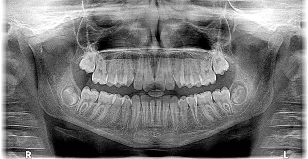
1. Radiography: X-ray radiography is the most widely used imaging technique. It involves passing X-rays through the body onto a digital detector or X-ray film. This technique helps diagnose fractures, lung conditions, dental issues, and other skeletal and soft tissue abnormalities.
2. Fluoroscopy: Fluoroscopy employs a continuous X-ray beam to create real-time moving images. It is often used during procedures such as angiography, where the flow of contrast agents through blood vessels can be observed.
3. Computed Tomography (CT): CT scans utilize X-rays to create detailed cross-sectional images of the body. CT scanners rotate around the patient, capturing multiple X-ray images from different angles. These images are reconstructed by computer algorithms, providing detailed three-dimensional information.
4. Mammography: X-ray mammography is an essential screening tool for breast cancer. Low-dose X-rays are used to capture images of the breast tissue, aiding in the early detection of abnormalities.
5. Interventional Procedures: X-rays are frequently used during interventional procedures, such as angioplasty or the placement of stents. They guide the physician by providing real-time imaging of the catheter's position and the targeted area.
Advancements and Safety Measures:
Over the years, advancements in X-ray technology have improved both imaging quality and patient safety. Some notable developments include:
1. Digital X-ray Systems: Traditional film-based X-ray imaging has largely been replaced by digital systems. Digital X-rays offer immediate image acquisition, higher image quality, and the ability to manipulate and store images electronically.
2. Reduced Radiation Dose: Efforts have been made to reduce radiation exposure during X-ray procedures. Modern X-ray machines employ techniques such as automatic exposure control and pulsed X-ray beams to minimize radiation dose while maintaining image quality.
3. Image-Guided Radiation Therapy (IGRT): IGRT combines X-ray imaging with radiation therapy, allowing precise targeting of tumors while sparing healthy tissues. It helps in adapting treatment plans and verifying the patient's position before each session.
4. Cone Beam CT: Cone Beam CT is a specialized form of CT imaging that provides high-resolution three-dimensional images with a lower radiation dose. It is commonly used in dental and orthopedic applications.
Safety measures are taken to
ensure that the benefits of X-ray imaging outweigh the associated risks. Protective lead aprons, collimators, and thyroid shields are used to minimize radiation exposure for patients and medical personnel. Furthermore, strict protocols and guidelines are followed to ensure appropriate and justified use of X-ray imaging.
Conclusion:
X-ray imaging has transformed healthcare by enabling non-invasive visualization of internal structures. From diagnosing fractures and dental issues to guiding complex interventional procedures, X-rays have become an indispensable tool in modern medicine. With continuous advancements in technology and a focus on patient safety, X-ray imaging continues to evolve, offering more precise and efficient diagnostic capabilities. As we look to the future, we can expect further breakthroughs in X-ray technology, enhancing our understanding of the human body and improving patient care.
About the Creator
Enjoyed the story? Support the Creator.
Subscribe for free to receive all their stories in your feed.
Comments
There are no comments for this story
Be the first to respond and start the conversation.