
Understanding Electrocardiography (ECG)
A Window into Heart Health
Introduction
The electrocardiogram (ECG or EKG) is a widely used medical tool that provides valuable insights into the electrical activity of the heart. It is a non-invasive procedure that has revolutionized the field of cardiology, aiding in the diagnosis and monitoring of various heart conditions. In this article, we will explore the fundamentals of an ECG, its significance in assessing heart health, and its role in diagnosing common cardiac abnormalities.
What is an ECG?
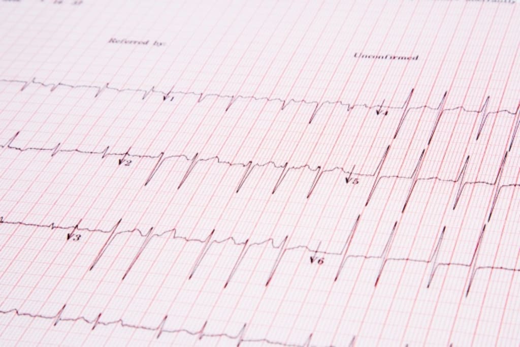
An electrocardiogram is a graphical representation of the electrical impulses that regulate the heart's rhythm. It records the electrical activity generated by the heart as it contracts and relaxes. The procedure involves placing electrodes on specific points of the body, which detect and transmit the electrical signals to the ECG machine for analysis.
Understanding the ECG Graph
The graph produced by an ECG consists of multiple waveforms that represent different phases of the heart's activity. The key components of the ECG graph include:
1. P Wave: Represents the electrical depolarization of the atria (upper chambers) as they contract to pump blood into the ventricles (lower chambers).
2. QRS Complex: Reflects the depolarization of the ventricles and the subsequent contraction of the heart muscle.
3. T Wave: Illustrates the repolarization of the ventricles as they prepare for the next cycle.
Significance of ECG in Assessing Heart Health
1. Diagnosing Arrhythmias: ECG is crucial in detecting irregular heart rhythms, known as arrhythmias. It helps identify abnormalities such as atrial fibrillation, ventricular tachycardia, and bradycardia, allowing healthcare professionals to initiate appropriate treatment strategies.
2. Detecting Ischemia and Heart Attacks: ECG aids in diagnosing conditions related to insufficient blood flow to the heart, such as myocardial ischemia and heart attacks (myocardial infarctions). It identifies characteristic changes in the ST segment, indicating compromised blood supply to the heart muscle.
3. Assessing Cardiac Hypertrophy: ECG can reveal abnormalities in the size and thickness of the heart chambers, providing insights into conditions like cardiac hypertrophy. This condition often occurs due to high blood pressure or other underlying cardiac disorders.
4. Monitoring Cardiac Function: ECG is widely used for routine monitoring of cardiac health. It helps assess the effectiveness of medications, the impact of lifestyle changes, and the progression of heart conditions over time.
Diagnosing Common Cardiac Abnormalities
1. Coronary Artery Disease: ECG assists in diagnosing coronary artery disease by identifying characteristic changes in the ST segment, indicative of reduced blood flow to the heart muscles.
2. Heart Failure: ECG can show abnormalities in the heart's electrical activity caused by heart failure. These changes help healthcare professionals determine the severity of the condition and formulate appropriate treatment plans.
3. Long QT Syndrome: ECG is crucial in identifying Long QT Syndrome, a congenital condition that affects the heart's electrical system, potentially leading to life-threatening arrhythmias.
Conclusion
The electrocardiogram is a powerful diagnostic tool that enables healthcare professionals to assess the electrical activity of the heart, diagnose various cardiac abnormalities, and monitor heart health over time. Its non-invasive nature and widespread availability make it an invaluable asset in the field of cardiology. By providing a window into the heart's rhythm, the ECG plays a significant role in improving patient outcomes and ensuring timely interventions for heart-related conditions.
About the Creator
Enjoyed the story? Support the Creator.
Subscribe for free to receive all their stories in your feed.
Comments
There are no comments for this story
Be the first to respond and start the conversation.