Biological Psychology
Some more parts of the Brain and some methods of studying

This is a continuation of the previous article subtitled 'The Brain'. This article is about more parts of the brain and various methods to study this important organ of the body. The brain is important in many ways and here's how some parts function as well as how we can study this awesome piece of work.
More parts of the brain
The cerebellum is very quick in modulation and regulation of movement. It makes for smooth movements not rigid. It also is for the learning of movements and other learnings such as the control of emotions.
The brain stem very purpose is for the body's vital processes, as temperature, pulse, respirations, and blood pressure.
The reticular formation is while looking under a microscope but not a larger system for arousal.
The midbrain is for coordination and regulation as well as for emotion and motivation. It also deals with a variety of health issues.
The neo-cortex is thin and wide and folded in six layers of cells and columns and designates things by tasks.
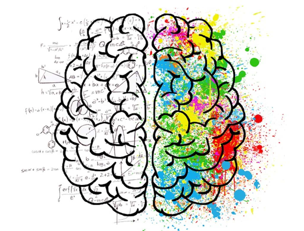
If you were to look at a sketch of the brain say looking down at the top of the brain you would see like down the middle and about halfway what is called the central fissure and this helps to delineate the parts to the brain. The front of the brain you will have two frontal lobes that initiate movement. Down the middle of the brain you will see the lateral fissure that will divide the brain in two it seems. Along the sides you will have two temporal lobes that are for judgment, analysis, and decision making. You also have two parietal lobes that are for higher processes, and finally you have the occipital lobe in the back of the brain and this is solely for vision.
_______________________
The brain helps deal with what is known as inhibition which is the suppressing of behavior or a strategy. The parietal lobes allow for touch for information from the skin, muscles, tendons, and joints. It is like a map for these locations. The temporal lobes allow for analysis of what to identify for vision. They are also used for memory and auditory means. This area is a complex system working together in a complex way.
It is a way that the MIND and BRAIN are together.
_________________________
Methods of studying the brain
The first way is through Gross anatomy that goes back as far as the Romans in brain study. This is to discern the structure of the brain. There is a lot of error and little clarity. In advancing to the microstructures attention will be to the tissues and for good optics of these you will need knowledge of the use of the microscope and chemistry.
From Germany the study of histology or the study of tissues and the cells that will make a map of the distribution of these cells. These maps were not very good at the time of the 1920's and 1930's. There are techniques known as ablation or better known as 'The Lesion Study' which is the removing of a portion of the brain to determine and to observe changes in function of the brain.
Brain research is still being done and one research project done in the 1950's were not that well done and not about the function or the structure of brain tissue. Electronics that are now used and available is of a bioelectrical nature of the brain as well for the patient are not put under and they are awake during these procedures. Electronics show very specific events. Processes are localized in the brain and a certain place in time. Localization before this with a person named Dax was able to show how the relationships between language and the left temporal lobe worked.
Phrenology is another form of localization and this study is attached to a person or from a record from a site or stimulated from a place with an electrodes that are placed on the brain or on the scalp and could be an early EEG (electroencephalogram) to see the net amount of action potentials of a large area and recorded. There are relatively few or a lot of relatively non-specific indicators of activity. The overall level of arousal of being awake, calm and preparing for sleep or just relaxing.
Modern measures of brain research are the use of CT scans (computed tomography) that measures structure and magnetic resonance (fMRI) measures activity function begins. There is not much difference between male and female, but the female will put more thought processes into the problems in different ways. Observable by function and changing very rapidly and conclusions are that the brain is always basically changing.
Functional analysis helps to understand the plasticity that can and does change the brain through aging. Adapt through the years and learning new skills that change life made a difference. Learning about different therapies that deal with brain damage and the various surgeries like the one Phineas Gage, the man who fell and ended up with a spike through his skull and survived. This is an example of how the brain changed through huge damage and can survive and what brain tissues recover from said damage. We may be able to restore and repair the brain.
In the development of the brain and with developmental engineering there are 100 billion trillion gial cells that must be connected properly. Things that are unproperly connected at times as in tastes and in music must work automatically a lot and wastes about one-half of the humans neurons where they are located or at the various connections. In the embryo there are three basic tissues called the skin, viscera , and neurons that still make 250,000 neurons a minute during the nine-month gestation of a human. The neural cells that are also parent cells of the dorsal nervous system spends a lot of energy and food even though very fragile during the process especially during the second trimester there are neural blasts that begin to migrate to where they should be in the body. They migrate by ameloid tendencies crawling along. Tissues may get lost on the way. The gial framework and cortex information or 'path-finding' to where neurons belong.
After reaching a place where they should be they 'aggregate' together to identify structure and function and differentiate between each other. In the adolescent and adult take on the characteristics of their roles as either senders or receivers they will now begin to specialize in transmissions that begin in the synapses and the validation of the cells. There is a selective cell death of 50% if not into a system. This is easily disruptive and changes will continue till 35 and above will continue to increase. Cell loss is inevitable and no new cells were formed in adults still have stem cells to learn new ways in the hippocampus. There is brain stimulation for new learning. This is an introduction to structural information of the neurons action potentials and the synapses in and out that follow these requirements.
(Please remember these articles are from my lecture notes from said course.) The next article will about how vision is seen from the brain.
About the Creator
Mark Graham
I am a person who really likes to read and write and to share what I learned with all my education. My page will mainly be book reviews and critiques of old and new books that I have read and will read. There will also be other bits, too.


Comments
There are no comments for this story
Be the first to respond and start the conversation.